Algorithms can make MRI scans sharper while subtly shifting clinical data
Machine learning sharpens scans and cuts examination times, but altering microscopic tissue measurements creates new calibration challenges for quantitative medicine.
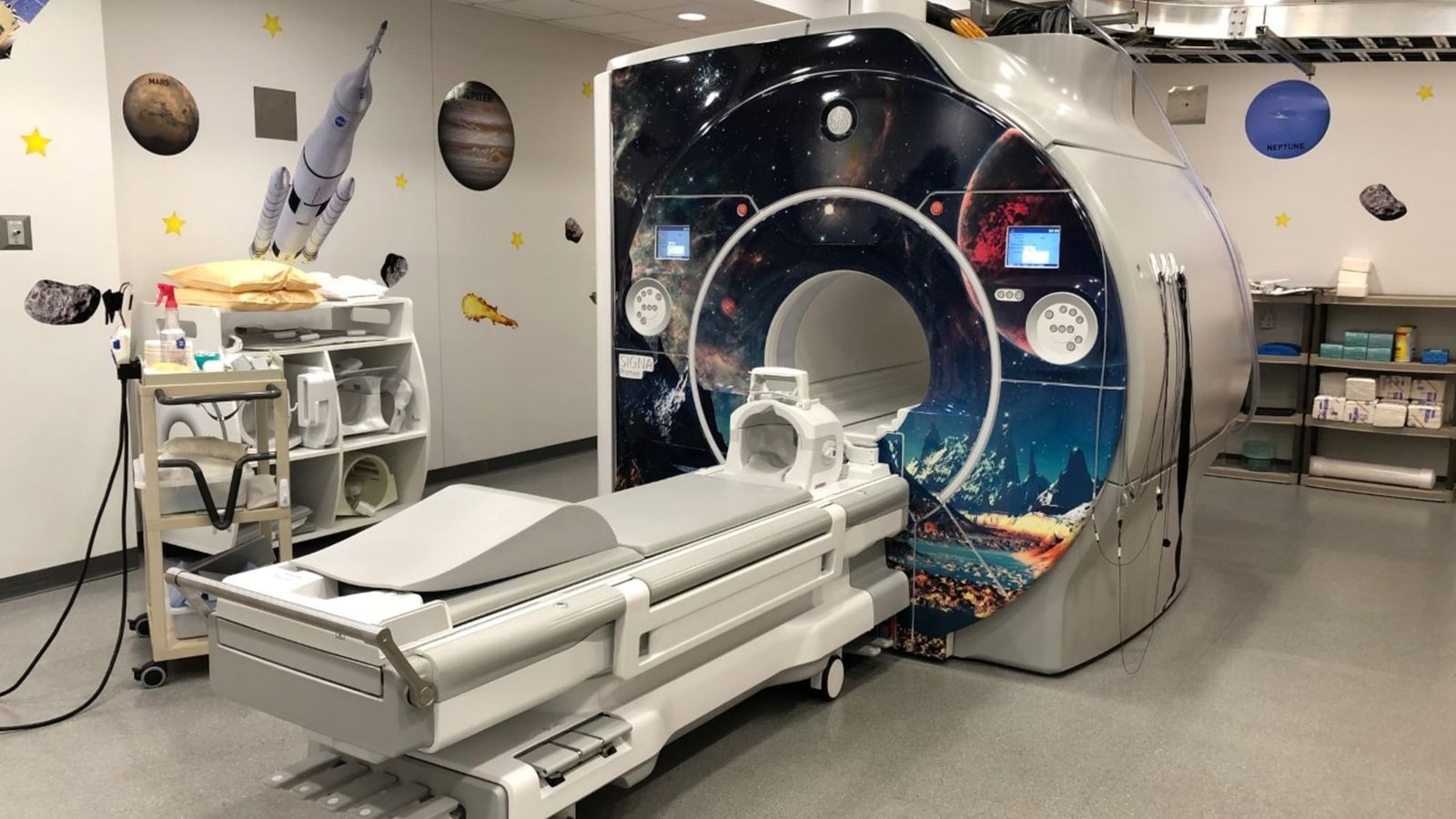
When a patient slides into a magnetic resonance imaging scanner, the resulting picture is often treated like a photograph of the inside of the human body. Clinicians and patients tend to assume that a sharper picture is automatically a more faithful representation of anatomy.1 For decades, acquiring that sharpness has demanded a frustrating clinical compromise. Securing fine anatomical resolution and detailed contrast requires scanners to record radiofrequency signals across extended periods.1 Longer sessions make it difficult for patients to remain still, and even slight physical movements introduce blurs that can obscure crucial tissue structures.1
Artificial intelligence offers a way to bypass that historical trade-off. Instead of gathering every raw radiofrequency point required to render a high-resolution view, a scanner can capture a smaller fraction of the data and rely on deep-learning models to reconstruct the unmeasured detail.1 Scans become faster, images look cleaner to the human eye, and clinics can evaluate more people in a single day.1 Yet sharpening an image through computation raises an essential question about the nature of the data: an algorithm that removes noise and fills in missing detail might make a scan look clearer, but it may also subtly alter the underlying measurements.
To understand why this alteration happens, consider how an imaging system translates physics into clinical pictures. The scanner does not take a snapshot. Instead, it places a patient within a strong magnetic field and registers radiofrequency signals emitted by hydrogen nuclei throughout the body.1 Those signals arrive as raw frequency values organized in an abstract mathematical framework called k-space.1 To convert those raw frequency numbers into an anatomical cross-section, a computer must execute a mathematical transformation.1 Computation has therefore always sat between the patient and the visible picture, but deep-learning systems dramatically expand how much interpretive work the software performs.
When an algorithm is tasked with completing a scan from sparse signals, it does not act as a neutral magnifying glass. It relies on learned statistical patterns to infer tissue boundaries, smooth out background irregularities, and guess what uncollected data should look like. In doing so, the software introduces its own mathematical tendencies into the resulting pixels. For a physician visually inspecting organs to identify a obvious fracture or a large lesion, that computational smoothing can provide valuable visual clarity. But modern radiology increasingly relies on computational tools that read the numbers behind those pixels rather than just their visual appearance.
How does deep learning change quantitative imaging?
Deep-learning reconstruction directly alters the numerical values of radiomic features that researchers extract from medical scans. As reported in an August 2026 study in Scientific Reports, applying deep-learning reconstruction algorithms to magnetic resonance scans significantly shifted the reproducibility and repeatability of these extracted computational markers.1 The investigation showed that while images produced through artificial intelligence appeared cleaner to visual inspection, the mathematical properties within the tissue regions changed when compared with conventional reconstructions.1
Radiomics is the practice of converting medical scans into extensive collections of quantitative data that describe tissue intensity, texture heterogeneity, and spatial patterns.1 Instead of relying exclusively on a radiologist visually evaluating whether a mass appears suspicious, analytical software calculates hundreds or thousands of numerical measurements across a suspected tumor.1 Scientists are actively testing whether these subtle mathematical profiles can predict how aggressively a tumor will grow, how tissue will respond to therapy, or whether a patient is at risk of disease recurrence.1
Because radiomic analysis depends on delicate differences in voxel intensity, changing the reconstruction software changes the quantitative outcome. The researchers in Scientific Reports demonstrated that reconstruction techniques matter fundamentally whenever scans are treated as scientific datasets rather than purely visual illustrations.1 An image processed through one neural network may look crisp and readable, yet its underlying numerical measurements may no longer match an image processed with standard methods or a different algorithm.1 The reconstruction model ceases to be an invisible tool and becomes an active component of the measurement system itself.1
Why does algorithmic variation complicate patient tracking?
Algorithmic variation creates a difficult calibration problem when clinicians attempt to track subtle biological changes over long periods. In standard laboratory medicine, a blood test is clinically actionable because measuring devices adhere to shared calibration baselines.1 If two different clinical laboratories analyze identical blood samples and report divergent figures, clinicians must determine whether the variance stems from the patient's physiology or from discrepancies between testing machines.1 Artificial intelligence introduces an identical calibration puzzle into medical imaging.
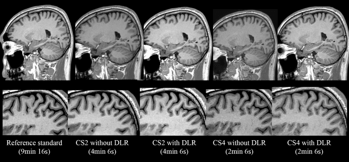
If two distinct deep-learning reconstruction models smooth microscopic tissue borders differently, the quantitative biomarkers pulled from their scans can deviate significantly.1 When that happens, clinicians cannot easily tell whether a shifting numerical score reflects true tumor shrinkage, disease advancement, or an artifact introduced by software. The physical scan itself remains subject to multiple technical variables, including magnetic-field strength, patient motion, acquisition protocols, slice thickness, contrast agents, and the specific sequence chosen by the technician.1 Layering deep-learning systems on top of those physical factors means small adjustments at any stage can ripple through downstream analyses.
This challenge grows more acute as patients undergo examinations across disparate facilities or explore private imaging options outside unified hospital systems. While patients understandably prioritize convenient appointments and rapid turnaround times, the long-term clinical utility of an examination often depends on technical consistency.1 Comparing scans taken months or years apart is one of medicine's most potent methods for catching disease progression early.1 If hospitals switch reconstruction software between appointments, meaningful comparisons become far more difficult to execute reliably.
What can current imaging algorithms not resolve?
Current research into deep-learning reconstruction cannot establish whether an individual algorithm will perform reliably across diverse clinical environments without extensive external testing. The findings reported in Scientific Reports highlight that algorithmic adjustments alter radiomic data, but they do not prove that artificial intelligence reconstruction is fundamentally flawed or clinically unsafe.1 Instead, they demonstrate that mathematical transformations alter numerical repeatability, leaving open the question of how specific diagnostic decisions might be affected across diverse hospital populations. Furthermore, the findings are observational measurements of feature variability, not proof that diagnostic accuracy invariably drops in routine care.
The field is simultaneously expanding into broader computational models. In July 2026, researchers reporting in Nature Medicine introduced NeuroVFM, a neuroimaging foundation model trained on more than five million three-dimensional magnetic resonance and computed tomography volumes gathered during routine medical care.1 Rather than training an isolated algorithm to recognize one specific illness, the model learned broad anatomical and pathological representations directly from large-scale clinical imaging, performing strongly across a range of diagnostic evaluations.1 Yet such foundation models also inherit the baseline characteristics of the scans they study.
For artificial intelligence to support long-term quantitative healthcare, the technical priorities of imaging research are shifting. Researchers are looking past visual polish to evaluate reproducibility, cross-scanner calibration, and stability across different patient demographics.1 The ultimate goal is not merely accelerating scanner hardware or generating visually striking pictures, but constructing integrated pipelines where acquisition, reconstruction, and machine learning function as a unified, mathematically reliable measurement system. Ensuring that algorithms faithfully reflect physical tissue remains the central requirement before these numerical techniques can direct widespread clinical care.
What this rests on
26 sentences trace to 1 source.
-
1
S2
Science 2.0 Document · science20.com · 12 Oct 2026 AI Is Making MRI Smarter: But It May Also Be Changing What We See See the source