Fossilized blood vessels reveal how T-Rex survived broken bones
Neutron imaging of a fractured rib from an ancient Tyrannosaurus rex shows preserved vascular networks that captured the animal healing before its death.
When heavy bones crack under stress, living tissue begins an immediate race to rebuild them. In the living body of an animal, microscopic channels carry blood, nutrients, and cellular repair crews directly to the fracture line to bridge broken structural surfaces. Most evidence of that delicate cellular activity disappears almost instantly upon death. Soft biological materials decay within days or weeks, long before heavy mineral sediments can replace decaying bone structures and turn ancient skeletons into rock.
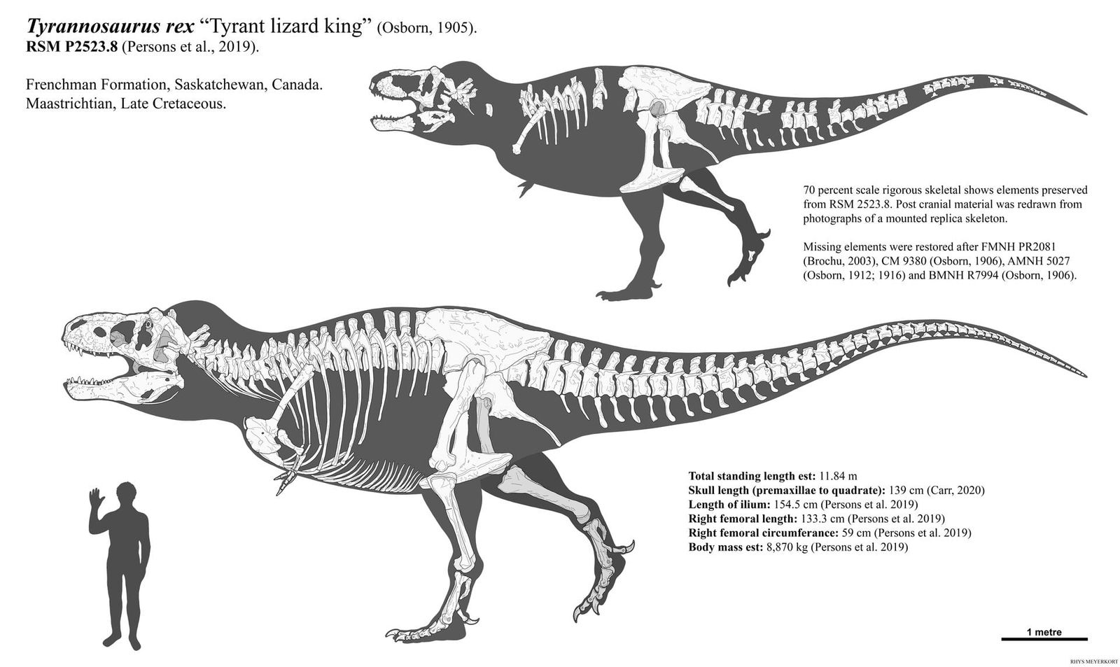
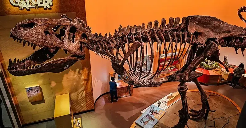
A deep look inside a fractured rib from an ancient carnivore shows that this cellular record does not always vanish. Researchers examining skeletal material from Scotty, the largest known Tyrannosaurus rex skeleton yet uncovered, have identified an intricate web of mineralized blood vessels preserved within a healed fracture.1 The preservation captures a transient biological response frozen in place 66 million years ago, showing how prehistoric animals sustained traumatic trauma and initiated internal skeletal repair.1
The findings, reported by physicists from the University of Regina working alongside specialists at the Department of Energy Oak Ridge National Laboratory, rely on beamline imaging techniques that examine the interior of dense fossils without slicing into them.1 By measuring how subatomic particles filter through dense crystalline fossils, the researchers managed to inspect fragile interior blood channels that normal radiation techniques often miss.1
How did soft tissue survive 66 million years of fossilization?
Scotty died and settled into a salty marsh before the broken rib could completely complete its healing cycle.1 That specific environmental setting drastically slowed normal microbial decomposition, giving surrounding dissolved minerals sufficient time to seep into the tiny biological channels before the tissue completely rotted away.1
The sequence began with a violent bone fracture sustained during the dinosaur's adult life. When the rib broke, iron-rich blood rushed into the injured site to establish new vascular structures that could deliver repair nutrients to knit the bone together.1 Before the internal repair work was finished, the animal perished in the saline wetland environment. The salt and lack of rapid decay allowed mineral deposits to stabilize and petrify the tiny vessel walls, turning what would normally be perishable biological tubes into permanent mineralized casts inside the solid fossil bone.
Mauricio Barbi, a professor of physics at the University of Regina in Saskatchewan, noted the rarity of finding such delicate structures inside a massive prehistoric skeleton.1 "It's like winning the lottery," Barbi said, pointing out that Scotty's rib contains a vast network of mineralized blood vessels that has never before been observed in a fossil.1 While paleontologists routinely study fossilized teeth and exterior bone morphology, intact soft tissue structures like internal vascular networks are almost always lost to decay.1
Why can subatomic particles detect structures that standard X-rays miss?
Neutron beams interact directly with atomic nuclei rather than electron clouds, allowing them to pass through dense heavy minerals while reacting with light elements such as hydrogen.1 Conventional X-ray imaging works in the opposite manner by scattering against dense, heavy elements, which makes traditional scans ideal for mapping solid bone walls while leaving soft tissue signatures faint or washed out.1
The technical investigation began in 2020 when Jerit Mitchell, then an undergraduate physics student at the University of Regina working under the direction of Barbi, detected initial signs of blood vessels inside Scotty's rib.1 Working at the Canadian Light Source, Mitchell used micro-computed tomography, a noninvasive X-ray scanning method that revealed fossilized soft tissue features on cut slices of the bone.1 The group subsequently paired those scans with synchrotron radiation and cellular microscopy at the Canadian Light Source to analyze the cellular structure around the healing fracture.1
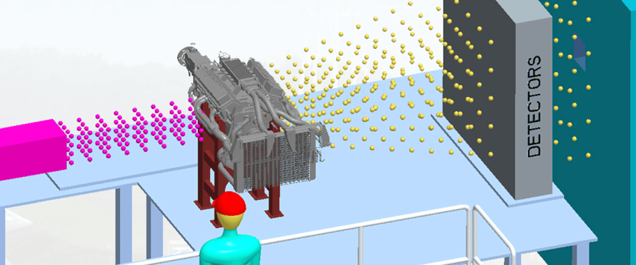
Those X-ray approaches proved that vascular structures were present, but they struggled to provide complete three-dimensional maps through thick, uncut fossil specimens. In April 2026, the investigation expanded to the Oak Ridge National Laboratory in Tennessee, where the team placed the fossilized bones into two complementary instruments: the MARS instrument at the High Flux Isotope Reactor and the VENUS instrument at the Spallation Neutron Source.1
Cold neutrons produced at the MARS instrument move at lower speeds and offer sensitive contrast across hydrogen-rich materials, making them suited for imaging fossilized scales, amber, and small skeletal fragments.1 High-energy neutrons generated at the VENUS facility penetrate deeply through dense matter without causing physical damage, allowing the researchers to generate high-resolution three-dimensional reconstructions of large bones like Scotty's rib.1 Marcella Berg, an assistant professor of physics at the University of Regina and a former postdoctoral researcher at Oak Ridge National Laboratory, noted that neutrons corroborated earlier synchrotron findings while mapping internal soft tissue properties without altering the physical specimen.1
What can these internal vascular images tell researchers about dinosaur biology?
Detailed three-dimensional maps of preserved blood vessels allow paleontologists to compare ancient bone reconstruction against the physiological repair mechanisms found in modern living animals. Because the fracture was actively healing when Scotty died, the density and orientation of the blood vessels illustrate how prehistoric reptiles marshaled circulatory resources to repair heavy structural skeletons.1
The analytical approach still faces clear technical boundaries. The scans provide spatial geometries of mineralized casts, but they cannot directly measure physiological metrics such as blood flow velocity, original chemical concentrations, or the exact timeline required for complete bone recovery. The data reflect mineralized structural replacements preserved under specific chemical conditions rather than living, flexible tissue. Furthermore, because intact vascular preservation requires an unusual burial environment, observations from a single specimen cannot demonstrate how typical or variable these healing speeds were across other predatory dinosaurs.
Field teams from the Royal Saskatchewan Museum originally discovered Scotty's skeleton in the Frenchman River Valley in Saskatchewan, a fossil deposit preserving ecological records immediately preceding the end-Cretaceous extinction.1 The researchers are now using the neutron beamlines at Oak Ridge National Laboratory to examine other fossilized dinosaur scales, bones, and amber samples from the region.1 By applying these noninvasive imaging methods across diverse specimens, the group seeks to determine whether similar vascular networks remain hidden inside other major museum fossil collections.1
What this rests on
23 sentences trace to 1 source.
-
1
N
Newswise Document · newswise.com ORNL Neutron Bone Imaging Brings T. Rex to Life See the source