Brain side matters in animal models of neurological disease
Preclinical studies that overlook differences between brain hemispheres risk misreading disease progression and drug efficacy, according to a systematic review.
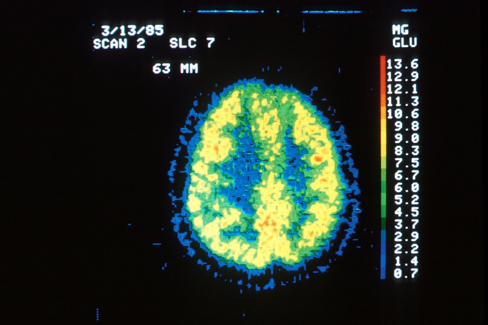
The mammalian brain looks symmetrically balanced at first glance, split into two mirrored halves across a central divide. Inside neural circuits, however, that symmetry breaks down. One hemisphere routinely handles tasks differently from the other, routing specific aspects of behaviour, memory, and spatial orientation through specialized physical pathways. When disease strikes, the two sides do not always experience the disruption equally.
For decades, medical researchers largely treated laboratory rodents as functionally balanced creatures unless a physical injury forced a difference. If an investigator tested a drug or modelled a disorder, they often assumed that measuring either hemisphere would yield an identical snapshot of the condition. Evidence has mounted that left and right hemispheres process metabolic demands, stress responses, and cellular changes along distinct tracks. If experimental models fail to register which side is affected, researchers risk misinterpreting how a disease develops and whether an experimental intervention actually works.
To understand why this imbalance matters, consider how a localized pathology interacts with the nervous system. Neural damage or genetic changes disrupt local signaling circuits. Because one hemisphere may express receptors differently or manage baseline neurotransmitter levels at varied concentrations, the disruption causes distinct functional deficits depending on which half takes the primary burden. Cells on one side can experience higher metabolic vulnerability, producing uneven behavioural symptoms in an animal. When scientists average measurements across both hemispheres or test only one side at random, those subtle differences wash out, clouding the underlying mechanism.
A systematic review published in Frontiers in Integrative Neuroscience by Maximilian Schuessler and Annakarina Mundorf brings this issue to the forefront. The authors evaluate how hemispheric asymmetries manifest across rodent models of neurological diseases, examining structural and physiological divergence between the brain halves.1 Their review collates evidence showing that rodent models display meaningful lateral differences in pathological conditions, challenging the standard assumption of bilateral equivalence in experimental design.
How did scientists discover asymmetry in mammalian brains?
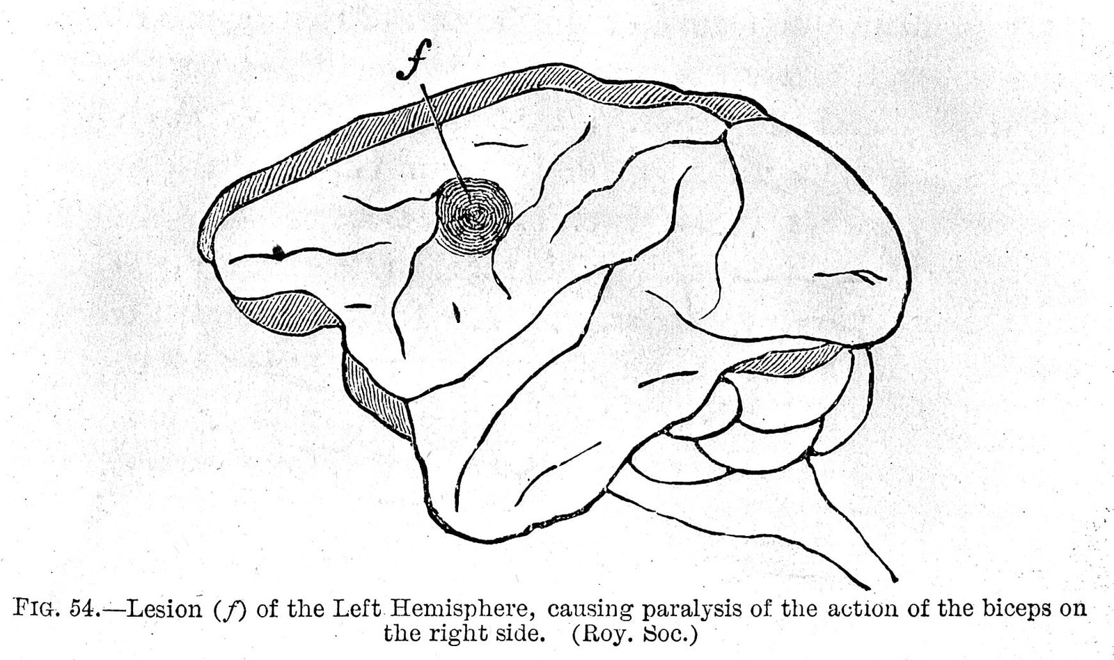
Pioneering clinical observations in humans first revealed that cerebral hemispheres divide labor rather than acting as interchangeable twins. Marc Dax and Paul Broca demonstrated in nineteenth-century studies that injuries localized to the left hemisphere produced severe speech impairments, establishing the principle of cerebral laterality.3 For generations, scientists regarded this asymmetry as a uniquely human feature tied to language. Later behavioral and anatomical surveys, however, identified lateralized traits across virtually all phylogenetic branches, demonstrating that brain asymmetry is an evolutionarily conserved feature across animals.3
Annakarina told Primary that splitting tasks between the two halves offers distinct evolutionary benefits. Hemispheric lateralization likely increases neural efficiency by allowing the two hemispheres to specialize in different functions, Annakarina said in response to questions from Primary, adding that this division of labor can improve processing speed, reduce redundancy, and enable multiple tasks to be performed simultaneously.contributed The evolutionary persistence across diverse taxa indicates that these functional advantages have been favored repeatedly over time.
Modern neuroimaging confirmed that functional and structural laterality extends far beyond speech, influencing memory, decision-making, and emotional processing.3 Researchers Madalena Esteves, Susana S. Lopes, Armando Almeida, Nuno Sousa, and Hugo Leite-Almeida previously documented that these lateral patterns remain plastic and responsive to environmental demands.contributed Their work noted that while adult human brains display widespread functional lateralization, laboratory rodents provide the vital experimental ground for unpicking the mechanistic causes behind these structural imbalances.3
Environmental shifts during development can also alter these neural patterns. Annakarina noted that broad environmental manipulations, such as zinc supplementation or prenatal exposure to toxins, can alter established patterns of hemispheric asymmetry.contributed Such findings indicate that lateralization is shaped by environmental influences during early growth as well as genetic programming, demonstrating substantial plasticity in asymmetric brain organization.
What did the systematic review evaluate in rodents?
Pre-existing functional dominance between the hemispheres helps determine how severely a neurological disease damages rodent neural tissue. Schuessler, based at the University Hospital Knappschaftskrankenhaus Bochum within Ruhr University Bochum, and Mundorf, based at MSH Medical School Hamburg, carried out a structured literature synthesis across published animal experiments.contributed Their analysis examined how different neurological conditions alter the two hemispheres, assessing where asymmetry appears in tissue damage, molecular signaling, and behavioral outcomes.
The biological drivers of this uneven vulnerability involve both pre-existing hemispheric traits and anatomical differences. Annakarina said that studies assessing hemispheric dominance before inducing pathology suggest that deficits are often more severe when the dominant hemisphere is affected.contributed Intrinsic hemispheric differences, including asymmetries in neurotransmitter systems, structural connectivity, or regional blood supply, may also contribute to differential vulnerability across the brain.contributed

The synthesis also addresses whether these lateral differences represent genuine biology or laboratory noise. Annakarina explained that if hemisphere of injury and hemispheric dominance had no consistent influence on outcomes across studies, then observed asymmetries would be far more likely to reflect experimental noise.contributed Because pre-existing hemispheric dominance consistently predicts the severity of impairments following experimentally induced damage, the observed divergences reflect biologically meaningful patterns.
What can these review findings tell researchers?
The conclusions of a systematic review reflect the published literature rather than a new laboratory experiment. Schuessler and Mundorf synthesized reported findings across disparate studies, which means their review cannot establish a single overarching biochemical mechanism for all hemispheric differences. Animal studies in the literature often vary in strain, sex, testing protocols, and surgical methods, introducing potential confounding factors across the reviewed corpus. The synthesis instead demonstrates an empirical pattern across multiple disease models, showing that hemispheric asymmetry is a regular feature rather than a rare anomaly.
These animal findings cannot be transferred uncritically into hospital settings. Rodent models provide valuable insights into the mechanisms and consequences of hemispheric asymmetries, but human neurological diseases are considerably more complex, Annakarina told Primary.contributed Annakarina emphasized that findings from animal models should therefore not be translated directly into clinical interventions, but rather highlight the importance of assessing the two hemispheres separately in human research and clinical practice.contributed
Accounting for brain laterality alters how preclinical animal experiments should be conducted in translational medicine. When investigators test new therapeutic compounds or evaluate disease mechanisms in rodents, tracking which hemisphere is affected could prevent false negatives and clarify inconsistent trial results. Unmasking these differences allows neuroscientists to match specific lateral vulnerabilities in animal models to the localized pathologies seen in human patients.
This piece was prepared from the paper in Frontiers in Integrative Neuroscience and public records together with answers from Annakarina to five questions from the Primary news team, completed September 2026.
What this rests on
14 sentences trace to 2 sources and 1 contributor.
-
1
F
Frontiers Document · frontiersin.org · 6 Aug 2026 Hemispheric asymmetries in rodent models of neurological diseases: a systematic review See the source
- 2 Contribution — Annakarina 12 statements added to this article
-
3
P
pubmed.ncbi.nlm.nih.gov Official record · pubmed.ncbi.nlm.nih.gov Unmasking the relevance of hemispheric asymmetries-Break on through (to the other side) - PubMed See the source
Article history
-
Published 18 Sep 2026, 18:14Assembled by the Primary desk from 2 sources · 1 contributor · 14 cited sentences
-
Annakarina
- Go to the sentence: The authors evaluate how hemispheric asymmetries manifest across rodent models of neurological diseases, examining structural and physiological divergence between the brain halves.
- Go to the sentence: Later behavioral and anatomical surveys, however, identified lateralized traits across virtually all phylogenetic branches, demonstrating that brain asymmetry is an evolutionarily conserved feature across animals.
- Go to the sentence: Hemispheric lateralization likely increases neural efficiency by allowing the two hemispheres to specialize in different functions, Annakarina said in response to questions from Primary, adding that this division of labor can improve processing speed, reduce redundancy, and enable multiple tasks to be performed simultaneously.
- Go to the sentence: Researchers Madalena Esteves, Susana S. Lopes, Armando Almeida, Nuno Sousa, and Hugo Leite-Almeida previously documented that these lateral patterns remain plastic and responsive to environmental demands.
- Go to the sentence: Their work noted that while adult human brains display widespread functional lateralization, laboratory rodents provide the vital experimental ground for unpicking the mechanistic causes behind these structural imbalances.
- Go to the sentence: Annakarina noted that broad environmental manipulations, such as zinc supplementation or prenatal exposure to toxins, can alter established patterns of hemispheric asymmetry.
- Go to the sentence: Schuessler, based at the University Hospital Knappschaftskrankenhaus Bochum within Ruhr University Bochum, and Mundorf, based at MSH Medical School Hamburg, carried out a structured literature synthesis across published animal experiments.
- Go to the sentence: Annakarina said that studies assessing hemispheric dominance before inducing pathology suggest that deficits are often more severe when the dominant hemisphere is affected.
- Go to the sentence: Intrinsic hemispheric differences, including asymmetries in neurotransmitter systems, structural connectivity, or regional blood supply, may also contribute to differential vulnerability across the brain.
- Go to the sentence: Annakarina explained that if hemisphere of injury and hemispheric dominance had no consistent influence on outcomes across studies, then observed asymmetries would be far more likely to reflect experimental noise.
- Go to the sentence: Rodent models provide valuable insights into the mechanisms and consequences of hemispheric asymmetries, but human neurological diseases are considerably more complex, Annakarina told Primary.
- Go to the sentence: Annakarina emphasized that findings from animal models should therefore not be translated directly into clinical interventions, but rather highlight the importance of assessing the two hemispheres separately in human research and clinical practice.