A three-minute brainwave test flags early risk of Alzheimer's disease
Passive electrical recordings of automatic visual recognition detected memory impairments linked to mild cognitive decline, offering a potential tool to identify patients who could benefit from emerging therapies.

Human memory often begins to deteriorate quietly, years before any clinical evaluation can detect noticeable changes in daily behaviour. In the earliest phases of cognitive decline, the brain may still compensate during conscious tasks, masking the progressive breakdown of underlying neural pathways. Traditional memory assessments rely on active participation, requiring an individual to listen to word lists, recall shapes, or solve puzzles under direct observation. That deliberate effort can produce test anxiety, and the resulting scores frequently fail to register the subtle functional deficits that emerge long before a formal diagnosis of dementia is made.
The delay between initial biological change and clinical confirmation creates a major barrier for treating neurodegenerative disorders. When memory problems finally become severe enough to interfere with independent living, substantial neural damage has typically already occurred throughout critical memory circuits. Early identification is especially valuable for individuals who notice minor lapses but continue to manage everyday routines without significant disruption.
Identifying that vulnerable window has become increasingly urgent with the arrival of a new class of medical therapies. Recently developed drugs designed to slow Alzheimer's disease, including lecanemab and donanemab, appear to deliver their greatest therapeutic benefit when administered at the earliest detectable stages of pathology.1 If medical teams cannot reliably distinguish which patients with mild symptoms are on a trajectory toward severe impairment, individuals who might benefit most from early intervention risk missing the narrow window where these treatments have their strongest effect.
Understanding how to detect that vulnerability without demanding strenuous effort requires looking at how the brain handles recognition automatically. When a person looks at an image, visual processing areas register the shape, color, and identity of the object in fractions of a second. If that object has been seen recently, the brain generates an immediate, unconscious electrical echo marking the stimulus as familiar. That automatic recognition response occurs without any conscious decision to remember or search through past thoughts. When early neurodegenerative disease begins to damage recognition networks, that electrical echo weakens, even while conscious recall remains superficially intact. By measuring electrical impulses at the scalp, sensors can record whether this automatic neural signal fires reliably or fades away.
A study published in the journal Brain Communications demonstrates that this passive electrical response can reveal memory impairment in just a few minutes.1 Researchers evaluated an electroencephalogram assessment known as Fastball, developed at the University of Bath in collaboration with the University of Bristol.1 The technique detected specific memory deficits among patients diagnosed with mild cognitive impairment, and identified individuals who face an elevated risk of progressing to Alzheimer's disease.1
Fastball tracks the speed and strength of automatic brainwaves while volunteers watch images flash across a monitor without requiring complex mental exercises.1 Small sensors placed across the scalp capture minute changes in electrical voltage produced by synchronised neural activity.1 By observing how the brain reacts to repeated visual cues, the system evaluates the health of recognition circuits without requiring the participant to answer questions or explain what they remember.
How can a three-minute brainwave recording capture early memory decline?
The Fastball test measures electrical activity across the scalp while a person watches a rapid stream of images on a screen, recording the brain's automatic reaction to pictures seen shortly before the session.1
Before the recording starts, researchers present participants with eight common images and ask them to name each item out loud.1 The participants receive no instruction to memorize the objects, nor are they warned to watch for them during the upcoming visual presentation.1 This design keeps the initial exposure entirely passive, preventing volunteers from adopting conscious memorisation strategies that might disguise natural memory deficits.
Once the initial naming step is complete, the participant sits before a monitor where hundreds of images appear in rapid succession over the course of three minutes.1 Each picture remains on the screen for a third of a second.1 Throughout that fast-moving stream, every fifth image is drawn from the set of eight pictures the participant named earlier.1 Scalp sensors continuously capture the brain's millisecond-by-millisecond electrical responses, allowing the system to isolate the automatic neural spike triggered whenever a previously viewed object reappears.
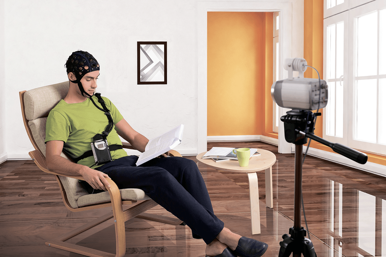
All of the test sessions in the study were conducted directly within participants' homes, avoiding the unfamiliar setting of a hospital laboratory.1 George Stothart, a cognitive neuroscientist at the University of Bath who developed the test, reported that administering the assessment at home was an essential element of the design.1 Conducting the evaluation in a familiar setting improves accessibility for older adults and helps reduce situational anxiety that frequently distorts performance on conventional clinical assessments.1
Why does distinguishing amnestic mild cognitive impairment matter?
Distinguishing amnestic mild cognitive impairment is essential because individuals affected by this condition face a far greater probability of developing Alzheimer's disease than patients with other forms of cognitive decline.1
The study included 54 healthy older adults alongside 52 individuals who had been diagnosed with mild cognitive impairment.1 People living with mild cognitive impairment experience measurable difficulties with memory, language, or executive thinking, but these challenges are not severe enough to prevent them from managing their daily activities independently.1 Because the condition encompasses a broad range of symptoms and underlying causes, some individuals remain stable for years, while others experience a steady progression toward dementia.
Within the clinical group, the researchers separated participants into those with amnestic impairment and those with non-amnestic impairment.1 Amnestic mild cognitive impairment primarily damages an individual's ability to retain and recognize memories for objects and events, reflecting dysfunction in memory structures closely tied to early Alzheimer's pathology.1 Patients diagnosed with non-amnestic impairment struggle with other cognitive domains, such as planning or verbal fluency, while their basic memory retention remains comparatively preserved.
The trial revealed that patients with amnestic mild cognitive impairment produced significantly reduced electrical responses during the Fastball test compared with healthy adults and participants with non-amnestic impairment.1 In the report, Stothart noted that this passive measure, built specifically for diagnosing Alzheimer's risk, proved sensitive to individuals who are at very high risk of decline but have not yet received a formal diagnosis of dementia.1 By pinpointing impaired recognition responses specific to amnestic cases, the test distinguished the patients whose clinical profiles place them at the highest statistical likelihood of worsening disease.1
What questions remain before passive testing enters clinics?
The Fastball test cannot determine with certainty which individuals will eventually develop Alzheimer's disease, operating as an indicator of elevated risk among people with mild symptoms.1
The findings represent an early evaluation in a modest group of 106 total participants, meaning the approach must be confirmed across much larger and more demographically diverse populations before it can be used in routine medical practice.1 The trial measured brainwave patterns at a single point in time, which means the current data cannot demonstrate how a person's electrical responses change as neurodegeneration progresses over several months or years.1 Memory impairment can also arise from numerous medical conditions outside of dementia, including vascular problems, sleep disorders, or metabolic imbalances.1 Consequently, researchers must determine how non-dementia health factors might alter scalp recordings and whether the test can accurately separate Alzheimer's pathology from other neurological disturbances.1
Evaluating those unresolved questions will be necessary to establish the clinical value of passive electroencephalograms. Vladimir Litvak, a researcher at the UCL Queen Square Institute of Neurology, indicated that the published trial represents an early step toward a practical clinical test.1 Litvak noted that a vital next objective will be determining whether the technology can reliably forecast how an individual's cognitive status will evolve over time and whether those measurements can help clinicians make informed treatment decisions.1
Earlier detection is increasingly seen as the foundation for modern dementia care. Julia Dudley, representing Alzheimer's Research UK, observed that new Alzheimer's treatments demonstrate greater efficacy when administered during the earliest stages of the condition, making early identification essential for patients to obtain maximum benefit.1 Dudley pointed out that future investigations must examine how passive brainwave assessments perform over extended periods, while exploring how the technique could function alongside existing diagnostic tools, including standard cognitive tests and blood tests, to build a reliable pathway for identifying those at risk.1
What this rests on
32 sentences trace to 1 source.
-
1
TG
The Guardian Document · theguardian.com Three-minute test helps identify people at greater risk of Alzheimer’s, trial finds See the source