Spheres of living cells and automated vision map the skin barrier
Testing topical compounds without animal tissue requires realistic human cellular shields, which spherical micro-tissues and machine vision can now track in living laboratory cultures.
The outermost shield of the human body maintains constant internal equilibrium while withstanding relentless environmental exposure. To protect tissues from dehydration, infectious microbes, and toxic chemicals, human skin continually rebuilds its protective surface through an orderly migration of cellular layers. Basal cells at the bottom of the epidermis divide, push upward, and change shape until they lock together into a dense, protective envelope that prevents fluids from leaking out and foreign agents from entering.
When this cellular defense falters, individuals suffer from chronic skin disorders, painful irritation, and systemic vulnerabilities. Developing medications to restore this protective envelope requires dependable methods to observe how human tissue assembles itself under controlled laboratory conditions. For decades, dermatological research has relied heavily on animal experimentation and flat cell cultures grown in plastic petri dishes, but both approaches present practical obstacles. Animal skin differs significantly from human skin in thickness and cellular behavior, while two-dimensional culture dishes force cells into unnatural flat configurations.
A study published on September 8, 2026, in the journal Frontiers in Bioengineering and Biotechnology presents an alternative experimental strategy.1 Lead author Tiziana Cesetti and colleagues across German academic institutes and biotechnology companies combined three-dimensional ball-shaped cell models with automated artificial intelligence to examine tissue maturation. The authors reported that coupling three-dimensional cellular spheres with deep-learning image analysis enables detailed observation of how specialized skin cells mature and construct a functional barrier.1
How does human skin construct its protective shield?
Natural skin relies on keratinocytes, the dominant cell type populating the epidermis, to execute an orderly developmental sequence known as differentiation. The process begins in the lower epidermal layer, where rounded basal keratinocytes divide continuously while resting on a supportive bed of dermal fibroblasts. As newly generated cells push toward the surface, they receive chemical signals and structural scaffolding from the underlying connective tissue, instructing them to cease division and begin their transformation.
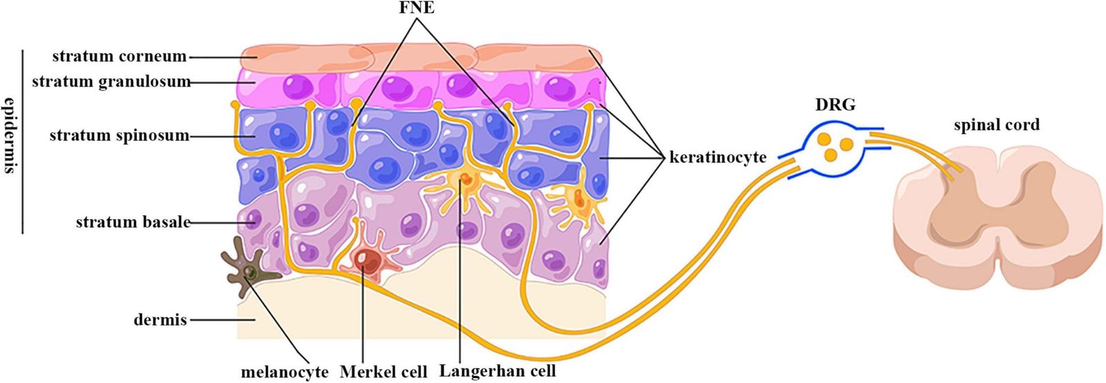
During this migration, the cells synthesise dense structural proteins, flatten their internal anatomy, and establish tight physical junctions with neighbouring cells. In the final phase of healthy tissue assembly, the cells discharge specialized extracellular lipids into the spaces between them, creating a mortar that seals the cellular brickwork. Finally, the cells lose their internal nuclei and form an impermeable outer stratum that prevents water loss and blocks external molecules. When cultured on conventional flat plastic, keratinocytes adhere directly to rigid synthetic surfaces without supporting fibroblasts, stalling before completing this terminal maturation.contributed
How do cell spheroids replicate the living epidermis?
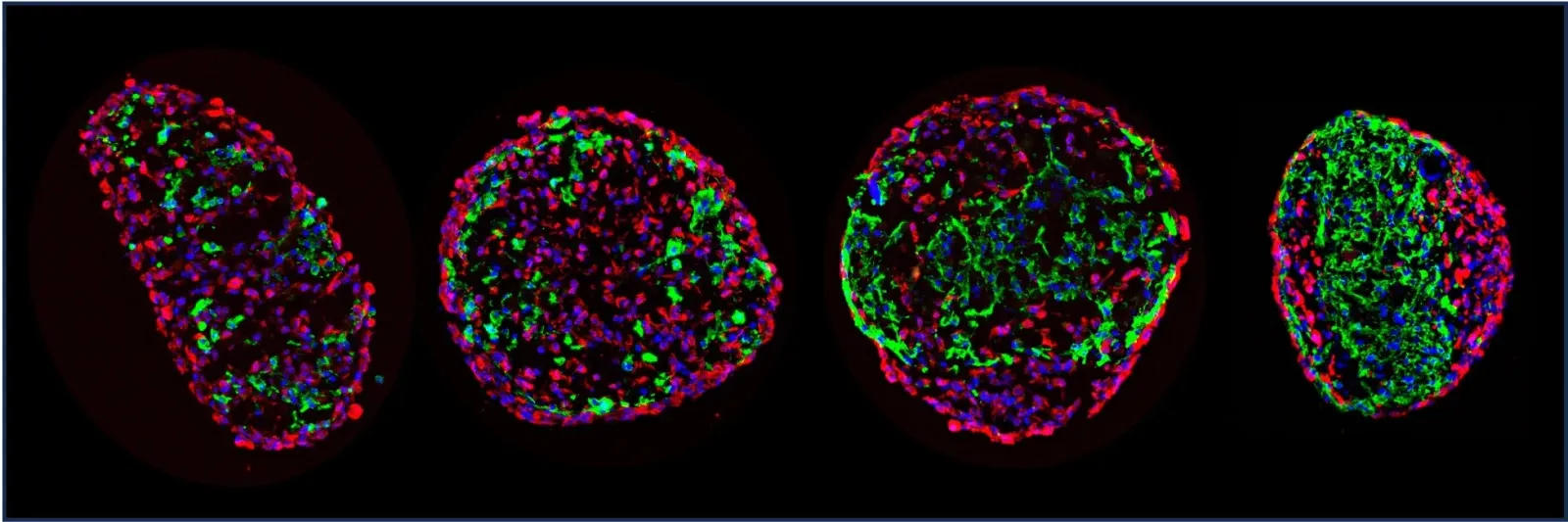
The experimental framework combines rounded clusters of living skin cells with deep-learning software designed to evaluate tissue structure from microscopic images.1 These spherical multicellular aggregates, termed spheroids, allow keratinocytes to self-organize in three dimensions, making physical contacts across all axes similar to natural tissue.1 Inside these microscopic spheres, the cells interact, polarize, and initiate differentiation pathways that mirror the organization of living epidermal layers more faithfully than flat cell monolayers.
The biological arrangement relies on a deliberate cellular pairing within each sphere. Tiziana explained in response to questions from Primary that each micro-skin spheroid contains a core of supportive connective cells enveloped by an outer mantle of surface cells.contributed Over approximately seven days, the keratinocytes organize into distinct developmental zones: round basal cells remain in direct physical contact with the internal fibroblasts, intermediate cell forms develop in the middle zone, and flat, fully differentiated cells seal the exterior surface.contributed Without fibroblasts providing extracellular matrix proteins and biochemical growth factors, this layered architecture fails to assemble.contributed
Tracking individual cells within these dense microscopic globes requires automated computational methods because manual visual inspection across hundreds of cell clusters is slow and subjective. According to the paper, the automated vision algorithms analyze high-resolution digital micrographs to measure structural indicators of keratinocyte differentiation and quantify how effectively the cellular spheres establish their protective seals.1 The researchers implemented Cellpose, an open-source deep-learning algorithm originally trained on diverse biological imagery across tens of thousands of segmented cell objects, to identify cell nuclei and monitor cellular shape changes from round spheres to elongated discs.3
The standard model required targeted adaptation to handle the complex optical conditions inside three-dimensional tissue clusters. Tiziana told Primary that while the pre-trained algorithm achieved respectable baseline results, it struggled when cells were tightly packed, dying, or shaped into elongated profiles characteristic of fully mature outer layers.contributed To overcome these tracking errors, human operators manually annotated corrected reference images, generating ground-truth data to retrain custom network weights that accurately tracked cell positions and protein distributions across the entire sphere.contributed
The research brought together teams across bioengineering, computational analysis, and clinical dermatology.1 Cesetti, Nathalie Couturier, Elina Nuernberg, Roman Bruch, Mario Vitacolonna, Mathias Hafner, and Rüdiger Rudolf conducted the investigation at Technische Hochschule Mannheim through its Institute of Molecular and Cell Biology and the CeMOS Research and Transfer Center. They collaborated with Claudia Buerger and Victoria Lang from the Department of Dermatology, Venereology and Allergology at Goethe University Frankfurt, Markus Reischl from the Karlsruhe Institute of Technology, and Torsten Fauth of BRAIN Biotech AG and Akribion Therapeutics GmbH.
Where does the automated vision system encounter limits?
The published research demonstrates a laboratory evaluation system without evaluating clinical treatments or diagnostic outcomes in human patients.1 As an original research article appearing in the biofabrication section of Frontiers in Bioengineering and Biotechnology, the work establishes an analytical workflow for cultured cells.1 Spheroids represent simplified cell aggregations that lack blood vessels, nerve endings, dermal papillae, melanocytes, and resident immune cells that populate native skin.contributed Furthermore, while human skin in living bodies requires two to four weeks to renew its outer stratum, the laboratory spheroids assemble their outer barrier in approximately seven days, demanding caution when extrapolating dynamic biological rates directly to human physiology.contributed
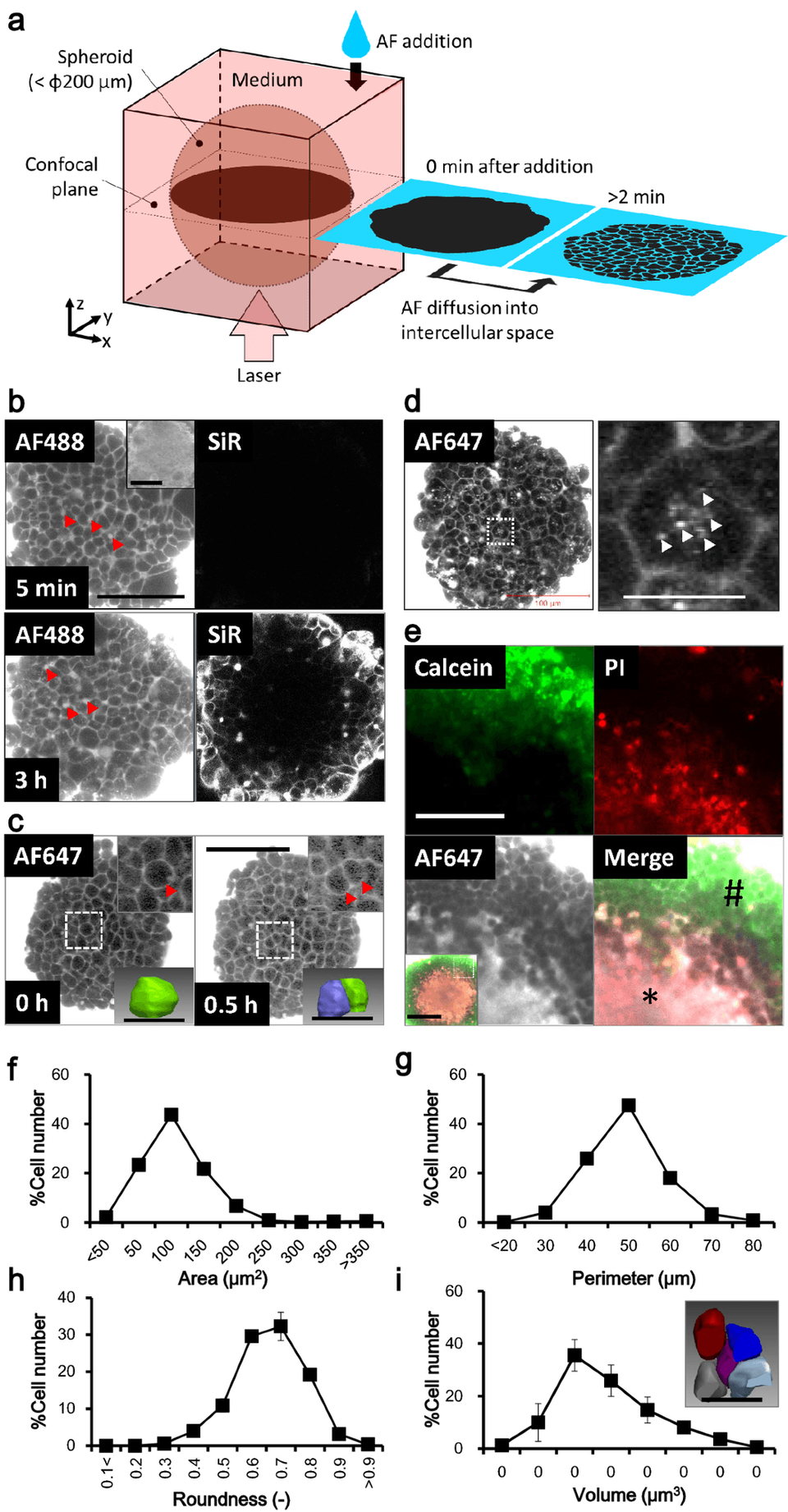
Assessing whether the resulting barrier truly stops foreign substances required specialized testing beyond visual protein markers. Standard laboratory tests calculate transepithelial electrical resistance across flat cellular sheets grown on porous membranes, but that electrical measurement cannot evaluate spherical geometries.contributed The researchers therefore developed a dynamic penetration assay using confocal live microscopy to follow the diffusion of a fluorescent hydrophilic dye.contributed In intact spheres, the fluorescent dye remained confined strictly to the outer cellular rim, penetrating the interior core only when chemical detergents intentionally disrupted the junctional seal.contributed
What can spherical tissue models reveal about topical drugs?
Quantitative analysis of dye diffusion permits sensitive measurement of how fast and how deeply test compounds penetrate tissue barriers under controlled conditions. Spheroid cultures produce a sealed surface barrier while remaining continuously submerged in liquid growth medium, contrasting with traditional organotypic air-liquid models that require weeks of atmospheric exposure.contributed However, confirming the structural maturity of this barrier requires future work measuring specific extracellular lipid classes, including ceramides, free fatty acids, and cholesterol, using analytical methods such as electron or Raman microscopy.contributed
The comparative experiments also revealed that barrier competence varies substantially between different donor sources. In head-to-head testing across several human keratinocyte lines, the most widely adopted standard cell line in dermatological research showed the weakest ability to construct an impermeable protective barrier.contributed Combining three-dimensional spherical co-cultures with deep-learning image analysis offers pharmaceutical researchers an objective tool to test compound absorption and evaluate experimental treatments for barrier-disrupting inflammatory disorders such as psoriasis and atopic dermatitis.contributed
What this rests on
24 sentences trace to 2 sources and 1 contributor.
-
1
F
Frontiers Document · frontiersin.org · 9 Sep 2026 Combining advanced 3D spheroid-based skin models with deep-learning-based image analysis enables in-depth investigation of keratinocyte differentiation and barrier function See the source
- 2 Contribution — Tiziana 21 statements added to this article
-
3
N
nature.com Paper · nature.com Cellpose: a generalist algorithm for cellular segmentation - Nature Methods Journal article See the source
Article history
-
Published 6 Oct 2026, 13:28Assembled by the Primary desk from 2 sources · 1 contributor · 24 cited sentences
-
Tiziana, lead author
- Go to the sentence: The authors reported that coupling three-dimensional cellular spheres with deep-learning image analysis enables detailed observation of how specialized skin cells mature and construct a functional barrier.
- Go to the sentence: When cultured on conventional flat plastic, keratinocytes adhere directly to rigid synthetic surfaces without supporting fibroblasts, stalling before completing this terminal maturation.
- Go to the sentence: The experimental framework combines rounded clusters of living skin cells with deep-learning software designed to evaluate tissue structure from microscopic images.
- Go to the sentence: These spherical multicellular aggregates, termed spheroids, allow keratinocytes to self-organize in three dimensions, making physical contacts across all axes similar to natural tissue.
- Go to the sentence: Tiziana explained in response to questions from Primary that each micro-skin spheroid contains a core of supportive connective cells enveloped by an outer mantle of surface cells.
- Go to the sentence: Over approximately seven days, the keratinocytes organize into distinct developmental zones: round basal cells remain in direct physical contact with the internal fibroblasts, intermediate cell forms develop in the middle zone, and flat, fully differentiated cells seal the exterior surface.
- Go to the sentence: Without fibroblasts providing extracellular matrix proteins and biochemical growth factors, this layered architecture fails to assemble.
- Go to the sentence: According to the paper, the automated vision algorithms analyze high-resolution digital micrographs to measure structural indicators of keratinocyte differentiation and quantify how effectively the cellular spheres establish their protective seals.
- Go to the sentence: The researchers implemented Cellpose, an open-source deep-learning algorithm originally trained on diverse biological imagery across tens of thousands of segmented cell objects, to identify cell nuclei and monitor cellular shape changes from round spheres to elongated discs.
- Go to the sentence: Tiziana told Primary that while the pre-trained algorithm achieved respectable baseline results, it struggled when cells were tightly packed, dying, or shaped into elongated profiles characteristic of fully mature outer layers.
- Go to the sentence: To overcome these tracking errors, human operators manually annotated corrected reference images, generating ground-truth data to retrain custom network weights that accurately tracked cell positions and protein distributions across the entire sphere.
- Go to the sentence: As an original research article appearing in the biofabrication section of Frontiers in Bioengineering and Biotechnology, the work establishes an analytical workflow for cultured cells.
- Go to the sentence: Spheroids represent simplified cell aggregations that lack blood vessels, nerve endings, dermal papillae, melanocytes, and resident immune cells that populate native skin.
- Go to the sentence: Furthermore, while human skin in living bodies requires two to four weeks to renew its outer stratum, the laboratory spheroids assemble their outer barrier in approximately seven days, demanding caution when extrapolating dynamic biological rates directly to human physiology.
- Go to the sentence: Standard laboratory tests calculate transepithelial electrical resistance across flat cellular sheets grown on porous membranes, but that electrical measurement cannot evaluate spherical geometries.
- Go to the sentence: The researchers therefore developed a dynamic penetration assay using confocal live microscopy to follow the diffusion of a fluorescent hydrophilic dye.
- Go to the sentence: In intact spheres, the fluorescent dye remained confined strictly to the outer cellular rim, penetrating the interior core only when chemical detergents intentionally disrupted the junctional seal.
- Go to the sentence: Spheroid cultures produce a sealed surface barrier while remaining continuously submerged in liquid growth medium, contrasting with traditional organotypic air-liquid models that require weeks of atmospheric exposure.
- Go to the sentence: However, confirming the structural maturity of this barrier requires future work measuring specific extracellular lipid classes, including ceramides, free fatty acids, and cholesterol, using analytical methods such as electron or Raman microscopy.
- Go to the sentence: In head-to-head testing across several human keratinocyte lines, the most widely adopted standard cell line in dermatological research showed the weakest ability to construct an impermeable protective barrier.
- Go to the sentence: Combining three-dimensional spherical co-cultures with deep-learning image analysis offers pharmaceutical researchers an objective tool to test compound absorption and evaluate experimental treatments for barrier-disrupting inflammatory disorders such as psoriasis and atopic dermatitis.