A single molecule quantum sensor measures tiny nuclear magnetic fields
Mapping individual proteins atom by atom could become possible using a single molecular radical to detect nearby nuclear spins through mechanical motion.
Determining the exact shape of an individual biological protein has long challenged structural biology, as conventional imaging methods require packing trillions of identical molecules into crystals or averaging signals across massive populations.12 When molecules are studied in bulk, subtle differences in how individual proteins twist, flex, and interact with other compounds vanish from view.
Quantum sensing offers a different path by using the properties of subatomic particles to measure external forces with high precision.12 Instead of observing molecules from afar, researchers aim to bring a magnetic probe directly alongside a single protein to record the unique magnetic signature of each atomic nucleus.23
A research team led by physics professor Raffi Budakian at the University of Waterloo has demonstrated that a single synthetic molecule can function as a nanoscale magnetic sensor.31 In a study published in Physical Review X, Budakian, doctoral researcher Sahand Tabatabaei, and their colleagues describe how an electron spin on a chemical radical detects nearby nuclear spins through minute mechanical vibrations.32
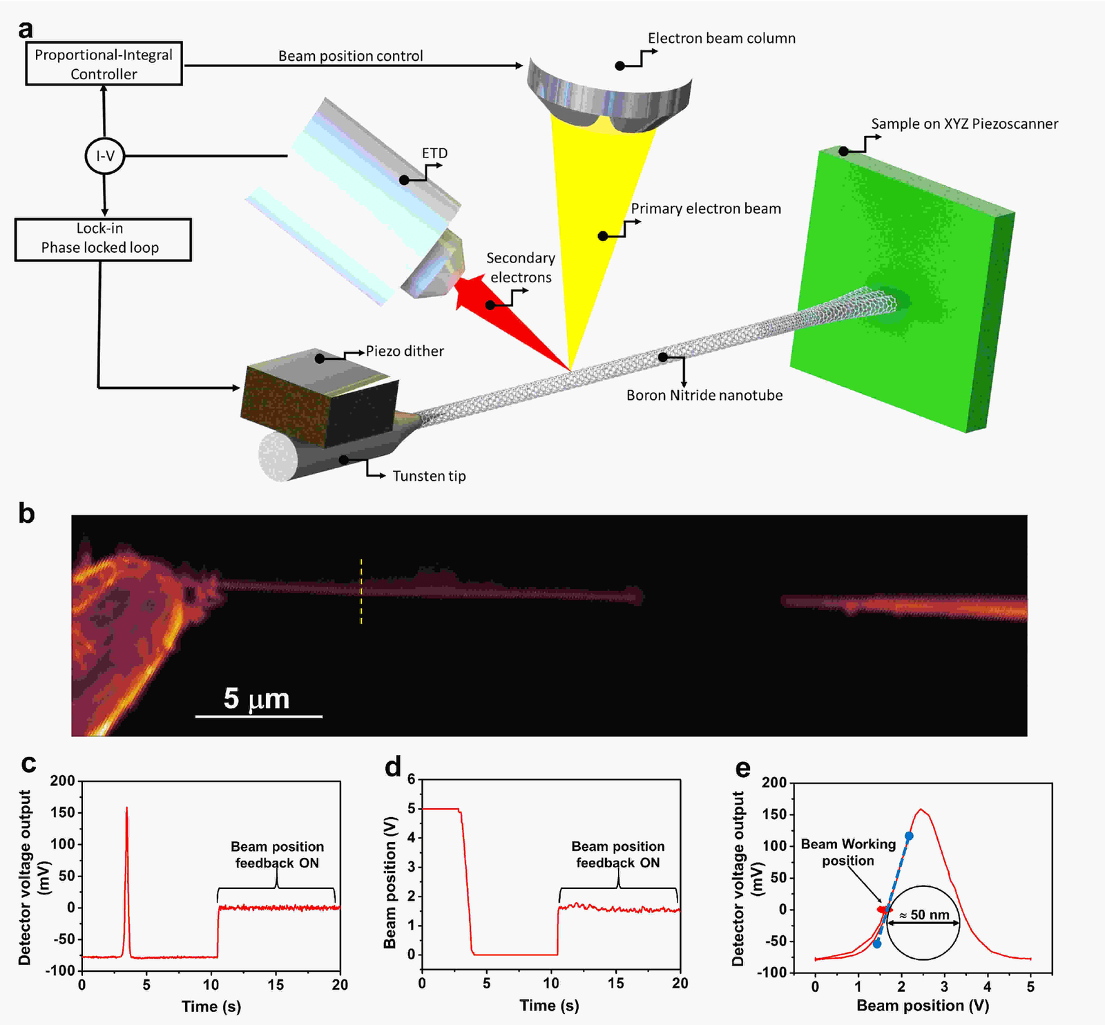
The sensing technique works by translating microscopic magnetic fields into physical motion.23 Many atomic nuclei possess an intrinsic quantum property known as spin, acting like miniature bar magnets.2 An unpaired electron on a neighboring molecule also carries spin, generating its own magnetic field.2 When an electron spin encounters nearby nuclear spins, its orientation wobbles at frequencies characteristic of those specific atoms.32 If shielded from environmental noise, that wobble persists long enough to be measured.31 To detect the shift without optical signals, the researchers attach the molecule to a microscopic flexible needle in a steep magnetic gradient.32 As the electron spin flips under microwave pulses, it exerts an oscillating physical push on the needle, converting magnetic resonance into mechanical motion.2
Why is mapping single biological molecules so difficult?
Conventional imaging techniques struggle to capture individual proteins because most tools require millions of identical copies packed into crystals or keep sensors locked inside rigid solids.21 X-ray crystallography requires orderly crystal lattices that many flexible proteins cannot form. Cryo-electron microscopy visualizes frozen samples, but reconstructing fine details still relies on computational averaging over thousands of distinct particle images.
Solid-state quantum sensors, such as nitrogen-vacancy centers in diamond, detect magnetic fields at nanometer scales but remain trapped within a host crystal lattice.2 Bringing a target protein within two or three nanometers of a diamond defect often destabilizes the sensor due to electric and magnetic surface noise.2 Because diamond sensors cannot easily bond to specific molecules, placing them close enough to resolve biological atoms has remained difficult.2
How does a single molecule act as a magnetic sensor?
A molecular sensor detects tiny magnetic fields through the orientation of its central unpaired electron, which shifts when nearby atomic nuclei exert a magnetic pull.3 For their sensor, the researchers selected trityl-OX063, a stable chemical radical with a narrow magnetic resonance line that makes it sensitive to local magnetic changes.21
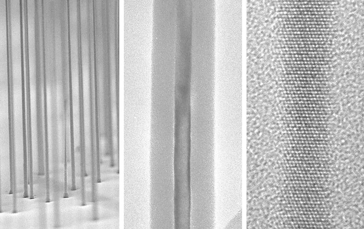
To read out the electron spin mechanically, the team fabricated a silicon nanowire oscillator measuring roughly 100 nanometers wide and 20 micrometers long.32 The researchers deposited a droplet of trityl-OX063 in a protective sugar mixture onto the tip of the nanowire.2 To prevent surface defects on the silicon from disrupting the spin, they mounted the droplet on a polystyrene spacer two to three micrometers long. That spacer increased the spin relaxation time to between 500 and 700 milliseconds, roughly a tenfold increase over configurations without a spacer.2
What keeps the fragile quantum signal alive?
A tailored sequence of microwave pulses keeps the molecular spin stable by cancelling unwanted magnetic disturbances from surrounding electrons.2 Left unshielded, electron spins in the sample lose their quantum coherence in 78 nanoseconds from environmental noise, or six microseconds under standard spin-echo pulses. The authors developed a modified dynamical decoupling sequence called XYXYd, which uses adiabatic microwave pulses to suppress electron-electron interactions across the sample.2
With this control sequence, the researchers extended the electron spin coherence time to about 400 microseconds, roughly 60 times longer than standard spin-echo techniques achieved in the same system.3 Operating in high vacuum at a base temperature of 4 Kelvin, the sensor performed nuclear magnetic resonance spectroscopy on hydrogen nuclei and carbon-13 nuclei.2 In its current setup, the device detected magnetic signals from about 10 nuclear spins in an attoliter droplet containing an estimated 140 sensor molecules.3
The current measurement demonstrates the operational physics of the sensor without producing a completed structural map of an unknown protein.32 The experiment detected small ensembles of spins in a sugar droplet under high vacuum at cryogenic temperatures, conditions far removed from the warm, liquid environment of living cells.2 The instrument also requires roughly a tenfold improvement in force sensitivity before it can isolate the signal of a solitary nuclear spin.32 Because the sensor spins were dispersed across an attoliter volume rather than tethered as a single radical to a biomolecule, true single-molecule imaging remains a future objective.
Chemical radicals provide a distinct advantage because chemists can synthesize and attach them directly to specific sites on proteins, placing the sensor at atomic proximity without the surface noise that afflicts diamond crystals.2 The Waterloo team aims to advance the mechanical readout toward single-spin detection, opening a path toward three-dimensional structural mapping of individual biological macromolecules to reveal how therapeutic drugs bind to disease targets.31
The record
31 sentences trace to 3 sources.
-
1
U
uwaterloo.ca Third-party reporting · uwaterloo.ca Using a molecule as a quantum sensor | Institute for Quantum Computing | University of Waterloo See the source
-
2
A
arxiv.org Paper · arxiv.org Long-Lived Mechanically-Detected Molecular Spins for Quantum Sensing Preprint · may not have been peer reviewed See the source
-
3
S
scientificinquirer Document · scientificinquirer.com · 13 Oct 2026 Researchers Turn a Single Molecule Into a Quantum Sensor That Could Map Proteins See the source