Misfolded immune defences can penetrate growing cells and disarm DNA repair
A newly identified class of cell-penetrating autoantibodies may explain how early childhood cancers emerge years before diagnosis by sequestering essential genome-maintenance proteins outside the nucleus.
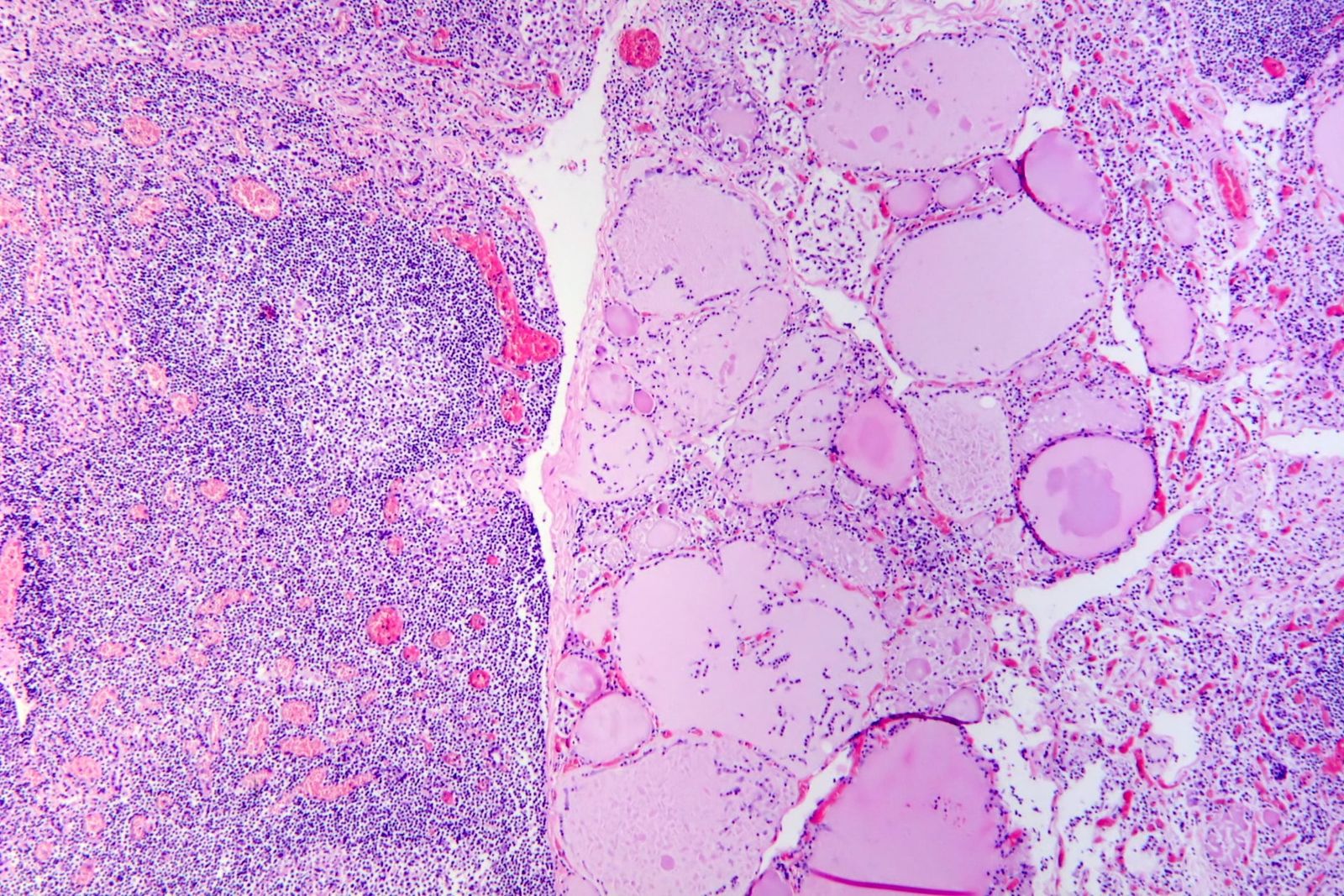
When adults develop malignant tumours, the underlying cause is frequently traceable to decades of wear and tear. Decades of cigarette smoke, ultraviolet sunlight, toxic chemical exposure, or simple chronological age introduce millions of random genetic typos into replicating tissues. In young children, that explanation fails entirely. Pediatric cancers strike when tissues are still forming, leaving little time for environmental hazards to inflict genomic injuries.12 Even more strangely, whole-genome sequencing reveals that pediatric tumours contain exceptionally few somatic mutations, while recognized inherited cancer syndromes account for only 6 percent to 10 percent of cases.1 For the vast majority of children with cancer, the root cause has remained unexplained.
Understanding why malignant cells arise in young bodies carries immediate clinical weight. An estimated 14,910 children and adolescents between birth and 19 years of age were expected to receive a cancer diagnosis in the United States in 2024.3 Although modern chemotherapy and supportive care have raised five-year survival rates past 85 percent across most pediatric cancers, treatment regimens remain physically grueling and can cause permanent secondary health difficulties.3 Pinpointing what initiates these rare diseases is the only path toward genuine prevention and earlier clinical detection.
Before examining experimental data, it helps to follow the chain of events that keeps normal tissue healthy. During rapid growth, human cells multiply continuously, copying billions of chemical letters in their genetic code. Physical strain and chemical metabolism inevitably fracture delicate DNA strands during this replication process. Under normal conditions, specialized housekeeping proteins immediately latch onto the damaged DNA strands, aligning the broken ends and stitching the sequence back together before division finishes. If an aberrant antibody enters the cell and traps those housekeeping proteins away from the nucleus, the broken strands remain unstitched. Mutations then begin to slip through uncorrected, accumulating across subsequent generations of cells until one lineage slips out of normal growth control.
A study published on October 14, 2026, in the journal Frontiers in Immunology by Anastassia Serguienko, Ola Myklebost, and colleagues at the University of Bergen in Norway suggests that such an autoimmune malfunction may occur years before cancer appears.43 The researchers found that children with cancer frequently harbor circulating autoantibodies, which are immune proteins that mistakenly bind the body's own normal tissues, aimed directly at critical DNA repair machinery.12 Crucially, the researchers detected these identical autoantibodies in archival blood collected from healthy children up to 15 years before those children were diagnosed with malignant tumours.31
Why do childhood cancers develop without decades of environmental damage?
Childhood cancers appear to develop when physiological DNA repair systems become functionally crippled by the body's own immune system, mimicking the effect of inherited genetic mutations.2 While adult tumours accumulate extensive damage from external mutagens over decades, pediatric cancers arise from primitive or developing progenitor cells during narrow developmental windows. When the internal molecular machinery that corrects ordinary replication slips is compromised, spontaneous errors cannot be repaired, accelerating malignant transformation in rapidly proliferating juvenile tissues.
To identify potential immune targets, the University of Bergen team conducted autoantibody profiling using high-density proteome microarrays, which are glass chips displaying thousands of individual human proteins to detect binding antibodies in liquid serum.5 The researchers screened serum samples collected at diagnosis from 175 cancer patients across nine distinct cancer types alongside 141 healthy control individuals of corresponding ages.13 The patient group included 84 pediatric patients and 91 adult patients with sarcomas and acute lymphoblastic leukemia, gathered from the Norwegian Sarcoma Study, the Norwegian Childhood Cancer Biobank, and the Oslo University Hospital Cancer Research Biobank.12
Gene set enrichment analysis revealed that autoantibodies targeting DNA repair pathways were enriched in patients aged 19 and younger, with a significance value of P = 6.4E-06 in young osteosarcoma patients, whereas adult cohorts showed no such pathway enrichment.1 A shared enrichment pattern appeared across pediatric patients with acute lymphoblastic leukemia, rhabdomyosarcoma, neuroblastoma, glioma, and Wilms tumour, reaching a significance value of P = 2.0E-04.1 Healthy children and older adult control subjects showed no comparable enrichment of these specific immune reactions.1
The discovery emerged unexpectedly from an effort to map tumour antigens. Myklebost said in response to questions from Primary that the project originally set out to identify disease-induced markers in connective tissue sarcomas, but "that finding was derailed by the very striking and surprising observation of these highly cancer-relevant antigens in children." The team dropped earlier priorities to investigate whether these immune responses might drive the initial development of childhood cancers.
The researchers defined a core set of autoantigens that appeared in at least 10 percent of patients across at least two separate cancer groups.1 Serguienko said in response to questions from Primary that this threshold was "an arbitrary threshold that, in our opinion, reflected a good combination of sensitivity and specificity to minimize bias." Among the most prevalent targets in pediatric patients were RAD52, a protein involved in repairing broken DNA strands through homologous recombination; NUMA1, an architectural protein supporting the mitotic spindle during cell division; and ALKBH7 and APBB1, two proteins involved in programmed cell death following unresolved DNA damage.1 In addition, the enzymes UNG and MGMT, which actively excise chemical lesions from DNA, were frequently targeted, alongside DDX58 and DHX58, which regulate innate immune responses.1
How can an antibody reach inside a cell to disrupt genetic maintenance?
Antibodies can physically cross the outer membranes of living human cells and accumulate around the cell nucleus to sequester repair machinery.13 Conventional immunological doctrine long held that antibodies function exclusively in the extracellular fluids and bloodstream, targeting invading pathogens or surface receptors while remaining excluded from the cell interior. The University of Bergen team tested whether circulating immunoglobulins from patient blood could enter intact cells and alter intracellular structures.
The biological mechanisms that permit large immunoglobulin molecules to enter living cells depend on specific structural features of their antigen-binding regions. In response to questions from Primary, Serguienko explained that entry depends on the variable region of immunoglobulin G, which means that only autoantibodies carrying particular amino acid sequences can penetrate living cells. Outside research into severe autoimmune disorders such as systemic lupus erythematosus has demonstrated that certain antibodies bind negatively charged sugar chains on the cell surface, enter through internalisation vesicles, and subsequently escape into the fluid interior, while others exploit surface nucleoside transporters or specialised membrane lipid microdomains.
The investigators applied donor serum diluted 1:40 in culture medium to cultured human osteosarcoma cells and primary human fibroblasts, incubating them at 37 degrees Celsius for two hours. Using high-resolution confocal microscopy, the researchers confirmed that immunoglobulin G antibodies from patient blood successfully crossed cell membranes. Once inside, antibodies from cancer patients formed distinct, dense cloud-like clusters located adjacent to the cell nucleus.31
Dual-staining experiments revealed that intracellular housekeeping proteins, including the repair factor RAD52, became physically concentrated within these cytoplasmic antibody clusters.32 By trapping essential repair molecules in the cytoplasm, the penetrating autoantibodies prevent them from entering the nucleus to resolve genomic lesions during cell division. Flow cytometry cell cycle analyses further demonstrated that this cellular penetration and clustering occurred most prominently during the phases when cells finish growing and prepare to divide their genetic material.12
What did pre-diagnostic blood samples reveal about future cancer risk?
Archived blood collected from healthy school-age children demonstrated that DNA repair autoantibodies were present up to 15 years before any clinical cancer diagnosis was made.13 To determine whether the antibodies preceded the appearance of tumours, the researchers accessed long-term longitudinal pediatric cohorts from the United Kingdom, specifically the Avon Longitudinal Study of Parents and Children and the Born in Bradford study.13
The investigators examined preserved serum from 30 healthy young participants who subsequently developed cancer before age 30 and compared them with 29 age-matched control participants from the same British cohorts who remained cancer-free.32 The archival blood samples had been drawn an average of about nine years before clinical tumour diagnosis, with individual intervals spanning between six and 15 years prior to detection.3 The authors noted that this multi-year interval corresponds closely with standard biological estimates of five to 20 years required for human malignancies to evolve from initial cellular damage.3
Statistical analysis using mixed-effects logistic regression models showed that 57 percent of the children who later developed cancer carried at least one of the core autoantibodies in their stored blood, compared with 14 percent of the matched healthy controls who never developed cancer.3 In pooled regression analyses, individuals carrying at least one core autoantibody were approximately eight times more likely to belong to the group later diagnosed with cancer, although the 95 percent confidence interval ranged widely from 2.5 times to 25 times due to the limited cohort size.32 When the six core targets were evaluated individually with false discovery rate corrections, autoantibodies targeting the repair enzyme UNG retained statistically significant elevation on their own.3
Addressing why only UNG met formal criteria after multiple-testing correction, Serguienko told Primary that anti-UNG was the most enriched autoantibody in patients. Serguienko observed that while statistical significance is a formal test of enrichment, other autoantibodies showed clear elevation in patient cohorts even when failing to clear that strict mathematical hurdle.
The long follow-up also captured people whose tumours appeared in early adulthood. Myklebost told Primary that autoantibodies detected in individuals diagnosed after age 20 represented only a subset of those seen in younger children, noting that UNG and MGMT were frequent in both groups. Myklebost observed that while sample sizes remain small, developing these malignancies by the mid-twenties suggests some underlying predisposition was involved.
What remains unproven about this autoimmune connection?
These findings describe a statistical association in human blood rather than definitive experimental proof that autoantibodies cause malignant disease.35 The study analyzed human serum samples and laboratory cell cultures, without conducting live animal experiments or interventional human clinical trials.3 Consequently, the researchers cannot exclude the alternative explanation that occult, microscopic pre-cancerous lesions had already formed years earlier and provoked an immune reaction as a secondary consequence.38
The statistical calculations also reflect real numerical limitations. The British pre-diagnostic group contained only 30 future cancer cases, and approximately half of those individuals were diagnosed after age 20 with adult-type malignancies.3 Furthermore, the presence of these autoantibodies is not absolute proof of future disease, as 14 percent of healthy control subjects in the pre-diagnostic cohort also tested positive for at least one core autoantigen.3
Myklebost cautioned that clinical interpretation must wait for broader epidemiological validation. In response to questions from Primary, Myklebost noted that researchers cannot interpret baseline positivity yet, explaining that "single positivities may not be sufficient, but multiple antibodies may increase the risk considerably." An exploratory check in adult leukemia patients with pre-cancer blood found no significant effect, which Myklebost said could indicate that the antibodies alter risk specifically during childhood development or that adult cases are dominated by different disease mechanisms.
Definitive causal proof will require animal testing. Myklebost told Primary that establishing whether these molecules initiate disease would require treating cancer-prone mouse pups during gestation and early life with cloned antibodies targeting relevant epitopes. If early cancer incidence fails to increase in treated animals under controlled conditions, that outcome would argue directly against the hypothesis.
Serguienko and Myklebost have filed a patent application covering the potential commercial use of these core autoantibodies as diagnostic biomarkers.3 No screening test based on this research exists for clinical practice today, and commercial diagnostic panels are neither approved nor recommended for healthy children. The American Cancer Society notes that common childhood cancer symptoms, such as persistent fevers, unexplained weight loss, unusual bruising, and localized bone pain, are far more likely to stem from routine childhood infections or minor injuries than from tumours.3 Further research within the University of Bergen KidImmune project, supported by the Trond Mohn Research Foundation, plans to evaluate these autoantibody markers in larger international patient registries and test whether purified patient antibodies directly induce DNA mutations in animal models.32
The record
49 sentences trace to 6 sources and 2 contributors.
-
1
F
Frontiers Document · frontiersin.org · 14 Oct 2026 Pathogenic autoantibodies targeting DNA repair proteins are shared by cancer patients and healthy children later diagnosed with cancer See the source
-
2
HT
Head Topics Third-party reporting · us.headtopics.com · 14 Oct 2026 Scientists identify novel autoimmune driver behind DNA repair breakdown in childhood cancer See the source
-
3
M
medicaldaily.com Third-party reporting · medicaldaily.com Antibodies Against DNA Repair Proteins Appeared Years Before Diagnosis in Some Children Who Later Developed Cancer See the source
-
4
F
-
5
F
Forskning.no Third-party reporting · forskning.no · 25 Oct 2026 Ny studie: Fant antistoffer i blodet opptil 15 år før barna fikk kreft See the source
- 6 Contribution — Ola Myklebost 7 statements added to this article
- 7 Contribution — Anastassia Serguienko 5 statements added to this article
-
8
N
Newsweek Third-party reporting · newsweek.com · 14 Oct 2026 Scientists Find Unusual Pattern in Children Later Diagnosed With Cancer See the source
Article history
-
Published 26 Oct 2026, 14:16Assembled by the Primary desk from 6 sources · 2 contributors · 49 cited sentences
-
Ola Myklebost
- Go to the sentence: Myklebost said in response to questions from Primary that the project originally set out to identify disease-induced markers in connective tissue sarcomas, but "that finding was derailed by the very striking and surprising observation of these highly cancer-relevant antigens in children."
- Go to the sentence: The team dropped earlier priorities to investigate whether these immune responses might drive the initial development of childhood cancers.
- Go to the sentence: Myklebost told Primary that autoantibodies detected in individuals diagnosed after age 20 represented only a subset of those seen in younger children, noting that UNG and MGMT were frequent in both groups.
- Go to the sentence: Myklebost observed that while sample sizes remain small, developing these malignancies by the mid-twenties suggests some underlying predisposition was involved.
- Go to the sentence: An exploratory check in adult leukemia patients with pre-cancer blood found no significant effect, which Myklebost said could indicate that the antibodies alter risk specifically during childhood development or that adult cases are dominated by different disease mechanisms.
- Go to the sentence: Myklebost told Primary that establishing whether these molecules initiate disease would require treating cancer-prone mouse pups during gestation and early life with cloned antibodies targeting relevant epitopes.
- Go to the sentence: If early cancer incidence fails to increase in treated animals under controlled conditions, that outcome would argue directly against the hypothesis.
-
Anastassia Serguienko, lead author
- Go to the sentence: Serguienko said in response to questions from Primary that this threshold was "an arbitrary threshold that, in our opinion, reflected a good combination of sensitivity and specificity to minimize bias."
- Go to the sentence: In response to questions from Primary, Serguienko explained that entry depends on the variable region of immunoglobulin G, which means that only autoantibodies carrying particular amino acid sequences can penetrate living cells.
- Go to the sentence: Outside research into severe autoimmune disorders such as systemic lupus erythematosus has demonstrated that certain antibodies bind negatively charged sugar chains on the cell surface, enter through internalisation vesicles, and subsequently escape into the fluid interior, while others exploit surface nucleoside transporters or specialised membrane lipid microdomains.
- Go to the sentence: Addressing why only UNG met formal criteria after multiple-testing correction, Serguienko told Primary that anti-UNG was the most enriched autoantibody in patients.
- Go to the sentence: Serguienko observed that while statistical significance is a formal test of enrichment, other autoantibodies showed clear elevation in patient cohorts even when failing to clear that strict mathematical hurdle.