Why colorectal cancer in young adults is proving so dangerous
Tumors in patients under 50 build protective fibrous barriers that disarm immune responses and deflect chemotherapy, according to a cross-age molecular atlas.
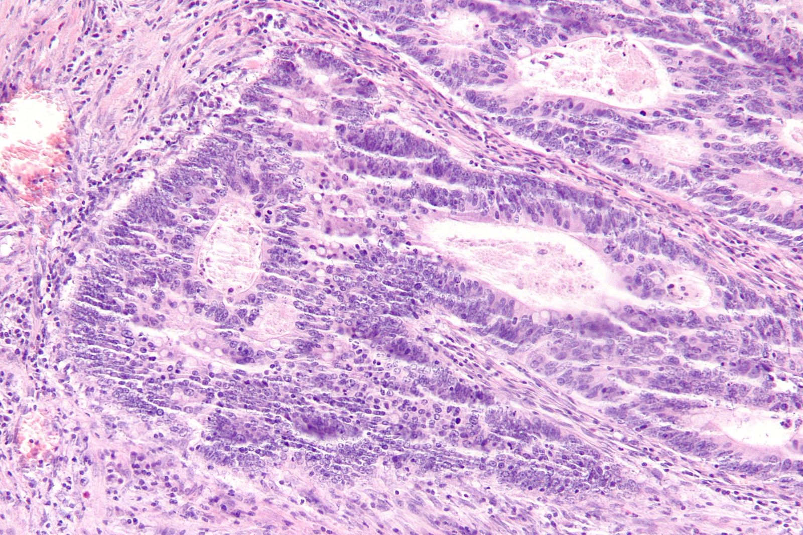
For decades, colorectal cancer was understood largely as a disease of aging bodies. The standard biological expectation was that decades of cell divisions and accumulated DNA insults eventually produced malignancies in the colon or rectum during retirement years. Since the mid-1990s, however, that clinical baseline has quietly inverted. Rates of colorectal malignancy have dropped steadily among older populations while climbing in adults under 50, appearing with unexpected frequency even in adolescents and young adults.12
These early-onset tumors do not simply arrive earlier in life; they behave with unusual clinical ferocity. Clinicians treating adults in their 20s, 30s, and 40s regularly encounter malignancies that present at late stages and resist standard cytotoxic drugs, defying the baseline assumption that younger, healthier patients with robust physiological reserve ought to tolerate therapy and survive longer. Early-onset colorectal malignancy has now become the leading cause of cancer death in the United States among people under 50.1
Understanding why these tumors kill requires tracing the biological sequence that normally allows therapies to clear an abdominal malignancy. First, an abnormal cell cluster in the colon lining begins to divide out of control. Second, surrounding immune sentinels detect foreign proteins displayed on the abnormal cells and recruit defensive killer cells to destroy the lesion. Third, when standard chemotherapy drugs arrive through the bloodstream, they must physically reach the cancerous cells and poison their division machinery before the cells spread. If any barrier shields the cancer from immune surveillance or physically prevents cytotoxic drugs from penetrating the growth, the tumor continues expanding and spreads into distant organs.
A study published on October 29, 2026, in the journal Nature Medicine identifies the structural differences that derail that therapeutic sequence in younger bodies.13 By comparing tumor specimens across the human lifespan, an investigation led by co-author Ana Anderson, a professor of neurology at Harvard Medical School and core faculty member at the Gene Lay Institute of Immunology and Inflammation at Brigham and Women's Hospital, alongside oncologist Kimmie Ng of the Dana-Farber Cancer Institute and gastroenterologist Ramnik Xavier of the Broad Institute and Mass General Brigham, established that tumors in young people build an entirely different internal microenvironment than those in older patients.14
What makes early-onset tumors structurally distinct?
Tumors in patients under 50 contain an unexpectedly high proportion of non-cancerous connective tissue cells relative to actual malignant cells.1 While older patients typically present with tumors dominated by proliferating epithelial cancer cells, tissue biopsies from younger patients show an inverted cellular landscape packed with dense structural stroma. In patients over 50, malignant cancer cells form the majority of the mass, whereas in younger individuals, connective tissue cells outnumber the malignant cells.1
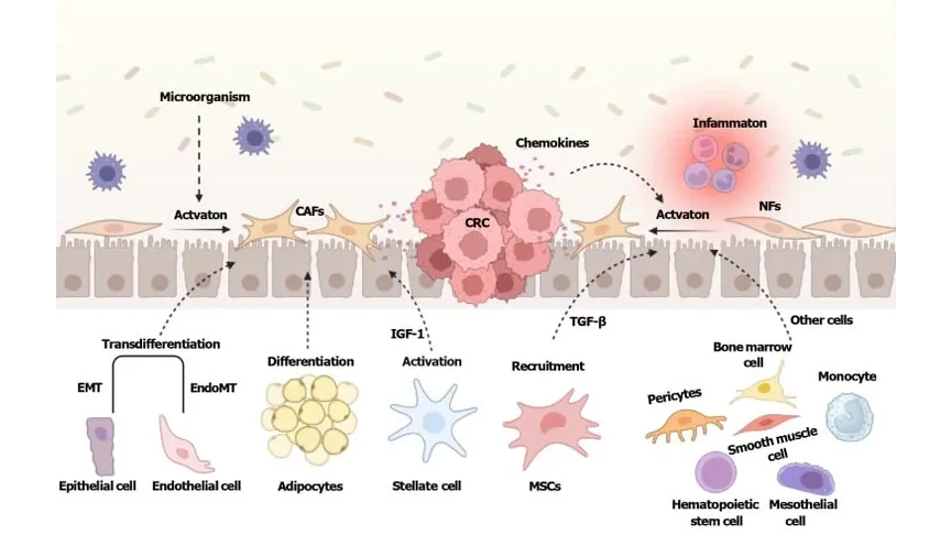
That abundance of connective tissue explains two major obstacles clinicians encounter when treating young patients. Rather than acting as harmless scaffolding, connective tissue cells secrete specialized biochemical compounds that actively suppress the local immune system.1 This chemical suppression disarms immune killer cells before they can target and destroy the malignant cells nearby.1 At the same time, the physical density of the connective tissue matrix creates a tight mechanical barrier that restricts the penetration of circulating chemotherapy molecules, blocking standard cancer drugs from ever reaching the cancer cells nested within.1
The structural changes also extend into the vascular network that feeds the growth. Earlier investigations led by Kosuke Matsuda, Satoko Ugai, and colleagues in the British Journal of Cancer demonstrated that tumors diagnosed at younger ages display lower overall vessel densities and marked reductions in specific vessel subtypes, including vessels defined by the endothelial marker CD34 and laminin beta 1.5 In their multivariable models, patients under 55 showed an odds ratio of 0.28 for high CD34-positive, laminin-beta-1-positive vessel density compared with patients aged 70 and older, pointing to distinct microenvironmental blood supply architecture in younger patients.5
How do young cancer cells move and evade treatment?
Malignant cells in younger patients demonstrate distinctive gene activity that governs cell-to-cell communication and accelerates migration.1 This altered signaling pattern enables individual cells to detach from the primary tumor bed and travel into surrounding tissues, creating a heightened risk of metastatic spread throughout the abdominal cavity and distant organs. In other cancer types, matching communication programs correlate closely with rapid metastatic colonization and clinical aggression.1
Younger tumors also exhibit marked shifts in non-coding genetic material. The analysis revealed an abundant presence of long noncoding RNA, which consists of molecular strands spanning more than 200 nucleotide building blocks that do not provide blueprints for building proteins.1 While coding messenger RNA directs cellular protein factories to construct enzymes and structural molecules, the exact physiological roles of these non-coding strands within colorectal tumors remain unresolved, presenting an unexplored molecular target inside early-onset disease.1
These transcriptional variations occur alongside widespread genomic instability and dampened immune cell engagement. An investigation published in npj Precision Oncology by Ting Peng and Qimin Zhan observed that early-onset colorectal cancers exhibit higher chromosomal copy number variation burdens than late-onset cases, with over 50 percent of tumors in patients under 40 classified as high copy number variation burden compared with under 35 percent in older cohorts.2 That analysis further demonstrated reduced proportions of tumor-infiltrating myeloid cells and weaker molecular interaction between tumor ligands and immune receptors in younger age categories.2
How did the researchers uncover these differences?
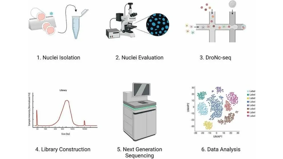
The research team mapped the cellular composition of tumors across decades of age by profiling single nuclei from frozen, banked surgical specimens. Using single-nucleus RNA sequencing, a laboratory technique that captures and measures active genetic transcripts inside preserved cell nuclei, the scientists evaluated 189 colorectal tumors, comprising 54 tumors from patients under 50 and 135 tumors from patients aged 50 and older, spanning patients from their 20s to over 90 years old.134
Analyzing archived frozen tissue allowed the investigators to examine specimens across a continuous age distribution rather than splitting patients into coarse, arbitrary bins.1 By assessing expression shifts across the age continuum, the group tracked subtle cellular trajectories that earlier comparison studies had obscured. The resulting atlas represents the largest high-resolution single-cell transcriptional database of colorectal tumors across the human lifespan.
The approach also avoided confounding variables that historically hampered cross-age research. By processing both younger and older specimens using a unified nuclear sequencing pipeline, the researchers distinguished authentic age-related microenvironmental phenotypes from technical artifacts. This revealed that the biological variance observed across age brackets reflects consistent physiological shifts within the tumor niche rather than accidental differences in surgical sampling or laboratory processing.
What questions remain unanswered?
The catalog of cellular differences in banked tissue cannot demonstrate that connective tissue expansion directly initiates the aggressive behavior seen in clinical wards. The findings represent observational transcriptional associations captured at the moment of surgical resection, leaving open the exact molecular mechanisms that prompt connective tissue cells to expand so aggressively in younger colons. Furthermore, tissue banking inevitably selects for patients who underwent surgical resection, meaning the cellular profiles may not fully capture the biology of unresectable, rapidly fatal primary presentations.
The underlying environmental and systemic causes driving the surge in early-onset colorectal cancer also remain unresolved by this single-cell atlas. While the study clarifies what young tumors look like once formed, it cannot identify which environmental exposures, dietary patterns, microbiome disruptions, or early-life chemical factors triggered the initial oncogenic process. Tracing the primary causes will require long-term prospective lifestyle and metabolic studies.
Translating these structural insights into improved clinical survival will require direct laboratory perturbation and prospective drug testing. Anderson and her collaborators are currently manipulating the long noncoding RNA molecules within cultured human cancer cells in petri dishes to test whether altering these transcripts slows metastatic migration or dismantles the connective tissue barrier.1 Identifying the molecular signals that prompt connective tissue to form protective shields may ultimately allow oncologists to dissolve those barriers, opening a path for existing chemotherapies and immune checkpoint inhibitors to reach early-onset cancers before they become fatal.
The record
20 sentences trace to 5 sources.
-
1
SP
Stephanie Pappas Document · livescience.com · 29 Oct 2026 Study reveals new clues to why colorectal cancer in young people is so deadly See the source
-
2
N
nature.com Paper · nature.com Single-cell integration analysis reveals molecular signatures of the tumor microenvironment in early-onset colorectal cancer - npj Precision Oncology Journal article See the source
- 3 Browse Articles | Nature Medicine Not published online
-
4
S
statnews.com Third-party reporting · statnews.com · 29 Oct 2026 New atlas of colorectal tumors sheds light on why cancers in young people are more dangerous See the source
-
5
N