A spliceosome flaw distorts the embryonic spine before bone forms
A new mouse model reveals how mutations in core splicing machinery leave physical embryo segmentation intact while silently disrupting the downstream genetic programs needed to build ribs and vertebrae.
During early embryonic development, the spine and rib cage take shape through a precise sequence of cellular events. Blocks of tissue called somites form in neat pairs along the developing back, establishing the primary physical blueprint for vertebrae and ribs. When this process stumbles, the consequences can alter the structure of the entire skeleton. In Cerebro-Costo-Mandibular Syndrome, children are born with severe axial skeletal abnormalities, including missing sections of their posterior ribs, an abnormally small jaw known as micrognathia, a cleft palate, and a bell-shaped chest cavity that often impairs breathing from birth.1 For years, the underlying developmental origin of these rib gaps remained difficult to explain because researchers could not study it with a disease-relevant animal model.
Understanding how these missing skeletal segments arise matters for pediatric genetics and fundamental developmental biology. Cerebro-Costo-Mandibular Syndrome is a rare congenital disorder linked to pathogenic variants in a single gene called SNRPB, which encodes a core component of the spliceosome.1 The spliceosome functions as the cell's internal molecular machinery for processing precursor messenger RNA, excising non-coding segments and joining protein-coding sequences together. Medical geneticists faced a longstanding puzzle: mutations in a ubiquitous, general-purpose splicing machine cause highly specific structural defects in the ribs and jaw rather than general cellular collapse. Because scientists lacked a disease-relevant animal model, testing targeted medical interventions or pinpointing the exact developmental window of vulnerability remained impossible.
The physical construction of the skeleton relies on a clear causal chain. Early embryonic precursor tissue first divides into segmented somites along the body axis. Inside each somite, molecular signals instruct cells on their precise anatomical identity, telling them whether to become muscular tissue, vertebral cartilage, or extending rib bone. Cells must then interpret those local biochemical cues, migrate and turn on expression of structural genes that deposit bone matrix. If the molecular instructions inside those nascent segments are scrambled, cells fail to differentiate and grow into mature bone structures even when the initial segmentation boundaries appear structurally intact.
A study posted on the bioRxiv preprint server indicates that the disorder's signature rib gaps stem from disrupted downstream programs that allow axial cells to execute their identity (migration, differentiation, patterning), rather than from a breakdown in somite formation or a loss of axial identity. Y. Dong, L. A. Jerome-Majewska, and colleagues at McGill University and the Research Institute of the McGill University Health Centre demonstrated that deleting exons two and three of the Snrpb gene in mice reproduces the full range of skeletal defects observed in human patients.1 Surprisingly, the embryonic somites in these mutant mice formed with entirely normal outer morphology and regular segmented patterning.1 The disruption occurred inside the cells at the level of gene expression, altering their developmental trajectory before bone formation ever began.
How did researchers reproduce the rare human syndrome in living tissue?
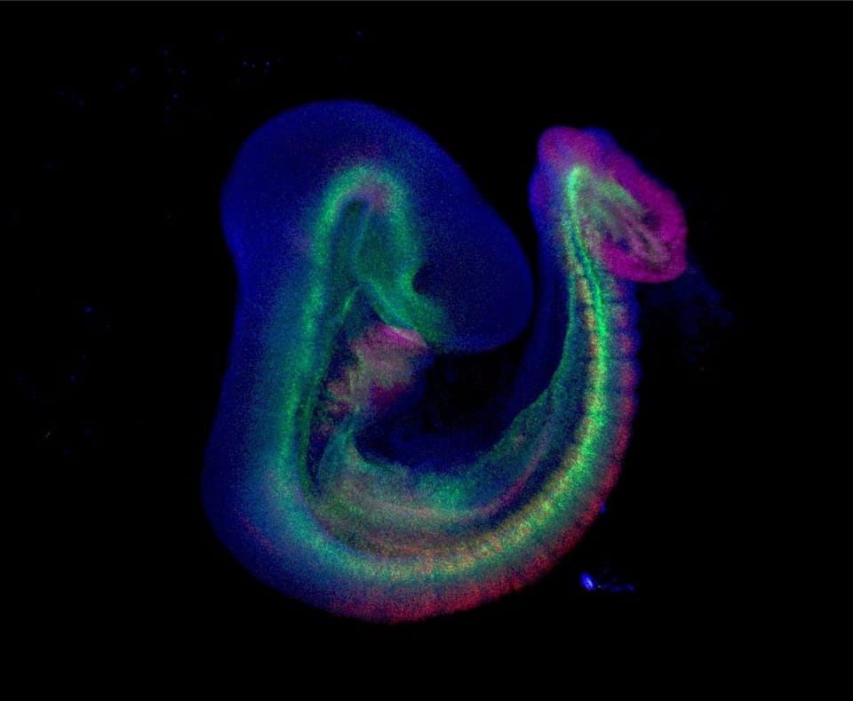
Researchers built an animal model for the syndrome by creating a conditional, mosaic heterozygous deletion of the mouse Snrpb gene using an inducible Cre-lox genetic switch. A previous model with ubiquitous heterozygous deletion of Snrpb caused early embryonic death, obscuring how later skeletal structures develop. By targeting exons two and three of Snrpb for mosaic heterozygous deletion specifically after gastrulation, the investigators allowed the embryos to progress past initial development.1 The resulting mutant mouse embryos exhibited the complete spectrum of human Cerebro-Costo-Mandibular Syndrome characteristics, including micrognathia, characteristic posterior rib gaps, and a markedly reduced thoracic cavity volume.1
The exact timing of this genetic deletion proved decisive in triggering the skeletal flaws. Loydie told Primary that the investigators did not directly measure the percentage of cells undergoing deletion, but noted that developmental timing dictated the outcome. Loydie said that when deletion was induced after embryonic day 8.5, just hours past their primary modeling window, rib and vertebral development appeared normal at embryonic day 14.5 despite mosaic deletion occurring across the embryo. "This suggests that the critical variable is not simply the proportion of cells deleted, but the developmental stage at which deletion occurs," Loydie said.
Why did physical segmentation form properly while bone development failed?
Physical segmentation of somites occurred normally because the initial structural boundaries of embryonic tissue segments operate upstream of the genetic programs that later execute skeletal differentiation. To uncover the molecular mechanism causing these missing skeletal segments, the team conducted transcriptomic profiling on mutant somites isolated at embryonic day 9.5.1 Gene expression analysis revealed widespread upregulation of genes in the p53 pathway, a cellular surveillance circuit that responds to stress, alongside mis-expression of components within the retinoic acid signaling network.1 This molecular signature suggested reduced retinoic acid signaling during the window when axial skeletal precursors are being patterned and begin to differentiate.
Furthermore, profiling identified aberrantly spliced transcripts among genes involved in post-transcriptional regulation and chromatin modification. Loydie told Primary that although somite formation, rostrocaudal polarity, and early sclerotome specification all proceed normally in mutant embryos, the cells ultimately fail to form proper vertebrae and ribs. "We propose that defective splicing does not erase axial cell identity but rather disrupts the downstream molecular programs that allow these cells to execute that identity: the gene regulatory networks governing cell migration, differentiation, and tissue patterning," Loydie said.
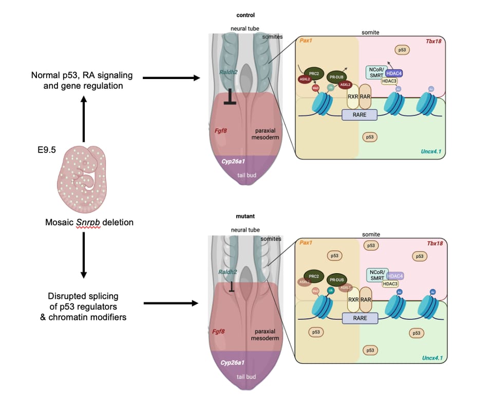
The initial appearance of the embryos concealed the underlying molecular damage. Loydie told Primary that when the team first observed normal somite morphology at embryonic day 9.5, they were struck by how subtle the early consequences were. Loydie explained that mutant embryos appeared morphologically indistinguishable from control specimens for several days, with morphological defects only becoming apparent at embryonic day 12.5. That observation prompted the researchers to examine the molecular changes occurring before physical deformities emerged, revealing the simultaneous disruption of p53 signaling, retinoic acid gradients, and chromatin regulation.
Why did retinoic acid supplementation fail to prevent the skeletal defects?
Dietary supplementation with retinoic acid failed to prevent the formation of rib gaps and jaw defects in the mutant mouse embryos because flooding the tissue with a single signaling molecule disrupted the delicate chemical balance across neighboring cells.1 The authors concluded that while reduced retinoic acid signaling is a defining feature of the diseased somites, it does not operate in isolation.1 Instead, concurrent activation of the p53 cellular stress cascade combined with aberrant chromatin modification contributes to the failure of skeletal specification.1
Loydie told Primary that identifying a disrupted pathway in one tissue does not mean whole-body supplementation will help. "In our case, supplementing retinoic acid across the whole embryo not only failed to rescue skeletal defects but exacerbated them, likely because the same signaling gradient that is disrupted in mutant cells must be precisely balanced in neighboring normal cells," Loydie said. Loydie added that therapeutic strategies for spliceosomopathies must account for interconnected pathways rather than targeting single disruptions in isolation.
These findings come from an animal model using an induced genetic switch, which carries inherent limitations. The results describe how conditional Snrpb deletion behaves in mouse embryonic tissue, but they cannot reproduce the exact mechanical and physiological environment of human gestation. The model relies on a mosaic heterozygous deletion of exons two and three, whereas patients carry heterozygous SNRPB variants (inherited or de novo), most of which cluster in the alternative exon 2 region and alter SNRPB splicing/expression rather than deleting exons and producing a null allele. Furthermore, the preprint has been posted to bioRxiv and has not yet completed formal peer review.
Resolving how these molecular disruptions interact will guide future work on potential therapies. Loydie told Primary that researchers cannot point to a single upstream trigger driving both p53 activation and retinoic acid failure, but favor a model where altered chromatin modifiers destabilize repressive gene states. Loydie noted that crossing mutant mice with p53 knockout strains could test whether cellular stress activation acts as the primary driver, while correcting chromatin modifier mis-splicing with antisense oligonucleotides could show whether chromatin defects sit atop the hierarchy. Those future experiments will help determine whether the cellular pathways act together or in sequence as the skeleton takes form.
The record
20 sentences trace to 1 source and 1 contributor.
-
1
DY
 Dong, Y., Bareke, E., Paquin, M., Smith, J., Alam, S. S., Majewski, J., Jerome-Majewska, L. A.
Paper
· biorxiv.org · 3 Sep 2026
Developmental pleiotropy revealed by mosaic heterozygous Snrpb deletion underlies CCMS-like axial skeletal defects
Preprint · may not have been peer reviewed
See the source
- 2 Contribution — Loydie 13 statements added to this article
Other consulted pages1 page
rqr-repro.org1 page
informatics.jax.org1 page
Article history
-
Published 18 Nov 2026, 18:08Assembled by the Primary desk from 1 source · 1 contributor · 20 cited sentences
-
Loydie
- Go to the sentence: Surprisingly, the embryonic somites in these mutant mice formed with entirely normal outer morphology and regular segmented patterning.
- Go to the sentence: Loydie told Primary that the investigators did not directly measure the percentage of cells undergoing deletion, but noted that developmental timing dictated the outcome.
- Go to the sentence: Loydie said that when deletion was induced after embryonic day 8.5, just hours past their primary modeling window, rib and vertebral development appeared normal at embryonic day 14.5 despite mosaic deletion occurring across the embryo.
- Go to the sentence: "This suggests that the critical variable is not simply the proportion of cells deleted, but the developmental stage at which deletion occurs," Loydie said.
- Go to the sentence: Gene expression analysis revealed widespread upregulation of genes in the p53 pathway, a cellular surveillance circuit that responds to stress, alongside mis-expression of components within the retinoic acid signaling network.
- Go to the sentence: Loydie told Primary that although somite formation, rostrocaudal polarity, and early sclerotome specification all proceed normally in mutant embryos, the cells ultimately fail to form proper vertebrae and ribs.
- Go to the sentence: "We propose that defective splicing does not erase axial cell identity but rather disrupts the downstream molecular programs that allow these cells to execute that identity: the gene regulatory networks governing cell migration, differentiation, and tissue patterning," Loydie said.
- Go to the sentence: Loydie told Primary that when the team first observed normal somite morphology at embryonic day 9.5, they were struck by how subtle the early consequences were.
- Go to the sentence: Dietary supplementation with retinoic acid failed to prevent the formation of rib gaps and jaw defects in the mutant mouse embryos because flooding the tissue with a single signaling molecule disrupted the delicate chemical balance across neighboring cells.
- Go to the sentence: Instead, concurrent activation of the p53 cellular stress cascade combined with aberrant chromatin modification contributes to the failure of skeletal specification.
- Go to the sentence: Loydie told Primary that identifying a disrupted pathway in one tissue does not mean whole-body supplementation will help.
- Go to the sentence: "In our case, supplementing retinoic acid across the whole embryo not only failed to rescue skeletal defects but exacerbated them, likely because the same signaling gradient that is disrupted in mutant cells must be precisely balanced in neighboring normal cells," Loydie said.
- Go to the sentence: Loydie told Primary that researchers cannot point to a single upstream trigger driving both p53 activation and retinoic acid failure, but favor a model where altered chromatin modifiers destabilize repressive gene states.